
Skip to content

Phương tiện truyền thông & Dung dịch phản ứng

40 products

Insulin, Human recombinant, Solution (5 mg/ml)

Volume: 5 ml Storage: +2°C to +8°C Sterility: Sterile-filtered Insulin Human Recombinant Solution is a chemically defined supplement commonly used for the cultivation of mammalian cell lines, including Chinese Hamster Ovary (CHO) cells. This cell culture–grade solution contains recombinant human insulin expressed in Saccharomyces cerevisiae, ensuring high purity and consistent performance in research applications. Application and Benefits Insulin is routinely added to serum-free and chemically defined media to promote cell growth and productivity. As a key regulatory hormone, insulin supports cellular uptake, utilization, and storage of glucose, amino acids, and fatty acids. It also inhibits the breakdown of glycogen, proteins, and lipids, thereby contributing to improved cell viability and metabolic stability in culture systems. The chemically defined formulation supports reproducibility and minimizes variability in sensitive cell culture workflows. Biological Properties and Usage Insulin is a two-chain polypeptide hormone naturally produced by the β-cells of the pancreatic islets. It has a molecular weight of approximately 5,800 Da. The α- and β-chains are linked by two interchain disulfide bonds, and the α-chain contains one intrachain disulfide bond. For cell culture applications, the solution should be handled under aseptic conditions and stored at +2°C to +8°C to maintain stability and performance. For research use only. Not for use in diagnostic or therapeutic procedures. Not for use in humans or animals.

€109.00

excl. tax and shipping

Insulin-Transferrin-Selenium (ITS), Solution 100x

Volume: 10 ml Storage: +2°C to +8°C Sterility: Sterile-filtered Insulin-Transferrin-Selenium (ITS) Solution (100x) is a chemically defined supplement designed for a wide range of cell culture applications. It is most commonly used as an additive to basal cell culture media to support cell growth under reduced-serum or serum-free conditions. Application and Benefits Our ITS supplement provides essential components required for the optimal performance of serum-free media. By supplementing conventional nutrient media with ITS, the fetal bovine serum (FBS) requirement for the routine maintenance of many cell lines can be significantly reduced. This helps minimize variability associated with serum use while maintaining consistent cell growth and viability. Insulin supports cellular uptake and metabolism of key nutrients, transferrin facilitates iron transport, and selenium contributes to antioxidant defense and enzymatic activity. Together, these components promote balanced cell metabolism and improved reproducibility in defined culture systems. Formulation and Usage Insulin-Transferrin-Selenium (ITS) is supplied as a 100x concentrated solution in Earle's Balanced Salt Solution (EBSS) without phenol red. For standard applications, dilute 1:100 into the appropriate basal medium to achieve the recommended working concentration. Store at +2°C to +8°C and handle under aseptic conditions to maintain product stability and sterility. For research use only. Not for use in diagnostic or therapeutic procedures. Not for use in humans or animals.

€59.00

excl. tax and shipping

McCoys 5A medium (modified), w: 3.0 g/L Glucose, w: stable Glutamine, w: 2.0 mM Sodium pyruvate, w: 2.2 g/L NaHCO3

McCoy's 5A Medium is a highly recommended and specialized medium designed to facilitate the growth and replication of viruses in primary cell cultures. It has gained significant recognition for its exceptional performance in various biological research applications. One prominent application of McCoy's 5A Medium is its utilization in the culturing of human colon carcinoma cell lines. Specifically, it has been employed in the study of the leucine-rich repeat-containing G-protein-coupled receptor (LGR5) and its role in the metastasis of colon cancer. This medium has been effectively employed in the cultivation of several colon carcinoma cell lines, including HCT116, RKO, FET, CBS, HCT116b, and TENN, enabling researchers to delve deeper into the mechanisms underlying colon cancer metastasis. In addition to its application in cancer research, McCoy's 5A Medium has proven to be indispensable in the study of osteoblasts. Researchers investigating the ion reactivity of calcium-deficient hydroxyapatite in standard cell culture media have utilized this medium to culture osteoblasts. This application has facilitated a better understanding of the interactions between osteoblasts and calcium-deficient hydroxyapatite, contributing to advancements in the field of bone research. McCoy's 5A Medium was meticulously formulated by modifying the amino acids found in Basal Medium Eagle to provide optimal support for liver tumor cells. This enriched formulation enables its suitability for a diverse range of established cell lines, as well as primary cells, further enhancing its versatility and applicability in various research settings. McCoy's 5A Medium extends its biochemical and physiological effects beyond liver tumor cells. It has been successfully employed to support growth in primary cultures of bone marrow, skin, gingiva, kidney, omentum, adrenal, lung, spleen, rat embryo, and other cell types. This wide range of applications attests to the broad utility of McCoy's 5A Medium in supporting the growth and maintenance of various cell types for comprehensive biological research. Formulation This McCoy's 5A medium (modified) contains 3.0 g/L of Glucose, stable Glutamine, 2.0 mM of Sodium pyruvate, and 2.2 g/L of NaHCO3. Quality control • pH = 7.2 +/- 0.02 at 20-25°C. • Each lot has been tested for sterility and absence of mycoplasma and bacteria. Maintenance • Keep refrigerated at +2°C to +8°C in the dark. Freezing and warming up to +37°C minimize the quality of the product. • Do not heat the medium to more than 37°C or use uncontrollable sources of heat (e.g., microwave appliances). • If only a part of the medium is to be used, remove this amount from the bottle and warm it up at room temperature. • Shelf life for any medium except for the basic medium is 6 to 8 weeks from the date of opening. Composition Components mg/L Inorganic SaltsCalcium chloride x 2H2O132.00 Magnesium sulfate97.67 Potassium chloride400.00 Sodium chloride6,460.00 Disodium hydrogen phosphate (anhydrous)504.00 Other ComponentsD(+)-Glucose (anhydrous)3,000.00 Glutathione (reduced)0.50 Meat Peptone600.00 Phenol red sodium salt11.00 Amino AcidsL-Alanine13.36 L-Arginine x HCl42.14 L-Asparagine x H2O45.03 L-Aspartic acid19.97 L-Cysteine x HCl x H2O31.75 L-Glutamine (stable)219.15 L-Glutamic acid22.07 Glycine7.51 L-Histidine x HCl x H2O20.96 L-Hydroxyproline19.67 L-Isoleucine39.36 L-Leucine39.36 L-Lysine x HCl36.54 L-Methionine14.92 L-Phenylalanine16.52 L-Proline17.27 L-Serine26.28 L-Threonine17.87 L-Tryptophan3.06 L-Tyrosine disodium salt26.10 L-Valine17.57 Vitaminsp-Aminobenzoic acid1.00 Ascorbic acid0.56 D(+)-Biotin0.20 D-Calcium pantothenate0.20 Choline chloride5.00 Folic acid10.00 myo-Inositol36.00 Nicotinamide0.50 Nicotinic acid0.50 Pyridoxal HCl0.50 Pyridoxine HCl0.50 Riboflavin0.20 Thiamine HCl0.20 Vitamin B122.00

€30.00

excl. tax and shipping

Medium 199, w: 2.7 mM stable Glutamine, w: 2.2 g/L NaHCO3, w: EBSS

Medium 199 is a versatile and widely used cell culture medium specially formulated for the cultivation of primary explants. This comprehensive medium combines essential vitamins, amino acids, and other factors to provide a fully defined nutrient source for a variety of cell types. It is particularly suitable for non-transformed cells, making it an invaluable tool for biological research. Medium 199 offers a range of applications in the field. It can effectively maintain the cumulus-oocyte complex (COC) and support the in vitro maturation of oocytes. Additionally, it is employed in the rinsing of aspiration lines during ovum collection from German Holstein cows. Moreover, Medium 199 serves as an excellent medium for the culture of cardiac endothelial cells derived from rats. These applications demonstrate the versatility and adaptability of Medium 199 to various experimental needs. History The development of Medium 199 in the 1950s marked a significant advancement in tissue culture media. Prior to its introduction, many culture media relied on animal-derived products and tissue extracts. However, Morgan and colleagues revolutionized the field by formulating a completely defined nutritional source for cell cultures. Through their experiments involving different combinations of vitamins, amino acids, and other factors, they discovered the exceptional growth-promoting properties of Medium 199. Quality control • pH = 7.2 +/- 0.02 at 20-25°C. • Each lot has been tested for sterility and absence of mycoplasma and bacteria. Maintenance • Keep refrigerated at +2°C to +8°C in the dark. Freezing and warming up to +37° C minimize the quality of the product. • Do not heat the medium to more than 37° C or use uncontrollable sources of heat (e.g., microwave appliances). • If only a part of the medium is to be used, remove this amount from the bottle and warm it up at room temperature. • Shelf life for any medium except for the basic medium is 8 weeks from the date of manufacture. Composition Components mg/L Inorganic Salts Calcium chloride x 2H2O 264,92 Iron (III) nitrate x 9H2O 0,72 Magnesium sulfate 97,67 Potassium chloride 400,00 Sodium acetat x 3H2O 82,95 Sodium chloride 6,800.00 Sodium dihydrogen phosphate x H2O 140,00 Other Components Adenine sulfate 10,00 AMP 0,20 ATP 1,00 Cholesterol 0,20 2‘-Deoxyribose 0,50 D(+)-Glucose anhydrous 1,000.00 Glutathione (red.) 0,05 Guanine x HCl 0,30 Hypoxanthine 0,30 Phenol red 10,00 D-Ribose 0,50 Thymine 0,30 Tween 80 4,90 Uracil 0,30 Xanthine 0,30 NaHCO3 2,200.00 Amino Acids L-Alanine 25,00 L-Arginine x HCl 70,00 L-Aspartic acid 30,00 L-Cysteine x HCl x H2O 0,10 L-Cystine 20,00 L-Glutamine stable 149,00 L-Glutamic acid 67,00 Glycine 50,00 L-Histidine x HCl x H2O 21,88 L-Hydroxyproline 10,00 L-Isoleucine 20,00 L-Leucine 60,00 L-Lysine x HCl 70,00 L-Methionine 15,00 L-Phenylalanine 25,00 L-Proline 40,00 L-Serine 25,00 L-Threonine 30,00 L-Tryptophan 10,00 L-Tyrosine 40,00 L-Valine 25,00 Vitamins 4-Amino benzoic acid 0,05 Ascorbic acid 0,05 D(+)-Biotin 0,01 Calciferol 0,10 D-Calcium pantothenate 0,01 Choline chloride 0,50 Folic acid 0,01 myo-Inositol 0,05 Menadione 0,01 Nicotinic acid 0.025 Nicotinamide 0.025 Pyridoxal x HCl 0.025 Pyridoxol x HCl 0.025 Riboflavin 0,01 DL-α-Tocopherol phosphate disodium salt 0,01 Thiamine x HCl 0,01 Vitamine A acetate 0,14

€40.00

excl. tax and shipping

NCI-H295R Cell Growth Medium

Discover the advanced NCI-H295R Cell Growth Medium, an expertly crafted nutrient solution specifically designed to foster the growth and maintenance of mammalian cell lines, including the specialized NCI-H295R cells. This medium stands out with its unique blend of Dulbecco's Modified Eagle Medium (DMEM) and Ham's F-12 (Nutrient Mixture F-12) in a precise 1:1 ratio, enriched with essential supplements to meet the intricate needs of cell culture. Enhanced Formulation for Superior Cell Culture Performance • Supplemented Baseline: The basic medium is already rich, combining DMEM's high amino acid and vitamin concentrations with the diverse nutrients of Ham's F-12. To this potent mix, we've added 5% Fetal Bovine Serum (FBS), providing crucial growth factors, hormones, and proteins. • Tailored Additives: Beyond FBS, this medium is fortified with critical supplements including Insulin, Transferrin, Bovine Serum Albumin, Selenious Acid, and Lineloic Acid. These additions cater to specific cellular requirements, promoting enhanced cell growth, metabolic function, and overall health. • L-Glutamine Boost: The presence of L-glutamine further amplifies the medium's efficacy, supporting essential metabolic processes and encouraging robust cell proliferation. Optimized for Cell Culture • pH Controlled Environment: Incorporating a bicarbonate buffer system and phenol red indicator, the medium ensures a stable pH environment. It is optimized for a 5% CO2 atmosphere, allowing for precise pH monitoring and adjustment. • Quality and Purity: Each lot of basic medium undergoes rigorous testing for sterility, mycoplasma, and bacteria, ensuring the medium's integrity. With a pH maintained medium, you can trust in the consistent quality and performance of this medium. Handling and Storage Maintain the medium's quality by storing it refrigerated at +2°C to +8°C, away from light. Avoid extremes, such as freezing or overheating, to preserve its efficacy. For partial use, gently warm the needed quantity at room temperature, ensuring the medium is brought to the appropriate condition without compromising its components. Shelf life With an 8-week shelf life from the manufacturing date, the NCI-H295R Cell Growth Medium is a reliable choice for your cell culture needs, ensuring the vitality and productivity of your research.

€150.00

excl. tax and shipping

PBS

Phosphate-buffered saline (PBS) is a widely used buffer solution in biological and chemical research. It plays a crucial role in maintaining the pH balance and osmolarity during various experimental procedures, including tissue processing and cell culture. Our PBS solution is meticulously formulated with high-purity ingredients to ensure stability and reliability in every experiment. The osmolarity and ion concentrations of our PBS closely mimic those of the human body, making it isotonic and non-toxic to most cells. Composition of Our PBS Solution Our PBS solution is a pH-adjusted blend of ultrapure-grade phosphate buffers and saline solutions. At a 1X working concentration, it contains: • 8000 mg/L Sodium chloride (NaCl) • 200 mg/L Potassium chloride (KCl) • 1150 mg/L Sodium phosphate dibasic anhydrous (Na2HPO4) • 200 mg/L Potassium phosphate monobasic anhydrous (KH2PO4) This composition ensures an optimal pH and ionic balance, suitable for a wide range of biological applications. Applications of Our PBS Solution Our PBS solution is ideal for various applications in biological research. Its isotonic and non-toxic properties make it suitable for substance dilution and cell container rinsing. PBS solutions containing EDTA are effective for disengaging attached and clumped cells. However, divalent metals such as zinc should not be added to PBS, as this can cause precipitation. In such cases, Good's buffers are recommended. Additionally, our PBS solution is an acceptable alternative to viral transport medium for the transport and storage of RNA viruses, including SARS-CoV-2. Quality Control • Sterile-filtered Storage and Shelf Life • Store at +2°C to +25°C, protected from light. • Once opened, store at 2°C to 25°C and use within 24 months. Shipping Conditions • Ambient temperature Maintenance • Keep refrigerated at +2°C to +8°C in the dark. Avoid freezing and frequent warming to +37°C, as it reduces product quality. • Do not heat the medium beyond 37°C or use uncontrolled heat sources such as microwave appliances. • If only part of the medium is to be used, remove the required amount and warm it to room temperature before use. Composition Category Components Concentration (mg/L) Salts Potassium chloride 200 Potassium phosphate monobasic anhydrous 200 Sodium chloride 8000 Sodium phosphate dibasic anhydrous 1150

€20.00

excl. tax and shipping

RPMI 1640, w: 2.0 mM stable Glutamine, w: 2.0 g/L NaHCO3

RPMI 1640 Medium, also known as RPMI medium, is a highly versatile cell culture medium widely utilized in biological research to cultivate various mammalian cells. Developed by George E. Moore, Robert E. Gerner, and H. Addison Franklin in 1966 at the renowned Roswell Park Comprehensive Cancer Center, this medium derived its name from its origin at the Roswell Park Memorial Institute (RPMI). Initially designed to support the growth of human leukemic cells in both suspension and monolayer cultures, RPMI 1640 Medium has evolved through modifications by researchers and commercial suppliers to become suitable for a diverse range of mammalian cells. It is exceptionally compatible with cell lines such as HeLa, Jurkat, MCF-7, PC12, PBMC, astrocytes, and carcinomas. RPMI 1640 Medium stands apart from other cell culture media due to its unique composition. It contains a substantial amount of phosphate, amino acids, and vitamins. Notably, it encompasses biotin, vitamin B12, and PABA, absent in Eagle's Minimal Essential Medium or Dulbecco's Modified Eagle Medium. Moreover, RPMI 1640 Medium exhibits significantly elevated concentrations of vitamins inositol and choline. However, it does not contain proteins, lipids, or growth factors. Consequently, supplementation with 10% Fetal Bovine Serum (FBS) is commonly required to provide optimal conditions for cell growth. The buffering system of RPMI 1640 Medium relies on sodium bicarbonate and necessitates a 5-10% CO2 environment to maintain a physiologically appropriate pH. The inclusion of the reducing agent glutathione further distinguishes this medium from others. Quality Control • Sterile-filtered Storage and Shelf Life • Store at +2°C to +8°C, protected from light. • Once opened, store at 4°C and use within 6–8 weeks. Shipping Conditions • Ambient temperature Maintenance • Keep refrigerated at +2°C to +8°C in the dark. Avoid freezing and frequent warming to +37°C, as it reduces product quality. • Do not heat the medium beyond 37°C or use uncontrolled heat sources such as microwave appliances. • If only part of the medium is to be used, remove the required amount and warm it to room temperature before use. Composition Category Components Concentration (mg/L) Amino Acids Glycine 10.00 L-Alanyl-L-Glutamine 434.40 L-Arginine 200.00 L-Asparagine H2O 56.82 L-Aspartic Acid 20.00 L-Cystine 2HCl 65.20 L-Glutamic Acid 20.00 L-Histidine HCl H2O 20.27 L-Hydroxy-L-Proline 20.00 L-Isoleucine 50.00 L-Leucine 50.00 L-Lysine HCl 40.00 L-Methionine 15.00 L-Phenylalanine 15.00 L-Proline 20.00 L-Serine 30.00 L-Threonine 20.00 L-Tryptophan 5.00 L-Tyrosine 2Na 2H2O 28.83 L-Valine 20.00 Vitamins p-Amino Benzoic Acid 1.00 D-Biotin 0.20 Choline Chloride 3.00 D-Calcium Pantothenate 0.25 Folic Acid 1.00 myo-Inositol 35.00 Nicotinamide 1.00 Pyridoxine HCl 1.00 Riboflavin 0.20 Thiamine HCl 1.00 Vitamin B12 0.005 Inorganic Salts Ca(NO3)2 4H2O 100.00 KCl 400.00 MgSO4 7H2O 100.00 NaCl 6000.00 NaHCO3 2000.00 Na2HPO4 800.00 Other Components D-Glucose 2000.00 L-Glutathione Reduced 1.00 Phenol Red Sodium Salt 5.30

€25.00

excl. tax and shipping

Stable Glutamin (200mM), L-Alanyl-L-Glutamine Solution

Volume: 100 ml Storage: ≤ –15°C Sterility: Sterile-filtered Stable Glutamine Solution (L-Alanyl-L-Glutamine, 200 mM) is a highly stable dipeptide form of L-glutamine designed as a direct replacement for conventional L-glutamine in cell culture media. L-glutamine is an essential amino acid and a major energy source for cultured cells, playing a critical role in cell growth, metabolism, and protein synthesis. Application and Benefits In standard liquid media, L-glutamine degrades relatively quickly at 37°C, leading to the formation of toxic byproducts such as ammonium ions that can negatively affect cell viability and experimental outcomes. Stable glutamine overcomes this limitation by providing a non-degrading dipeptide form that remains intact in culture conditions. Cells enzymatically cleave the dipeptide bond to release L-glutamine as needed, ensuring a continuous fresh supply while preventing the accumulation of harmful waste products. This makes the solution particularly advantageous for long-term cell cultures and high-density growth systems. Formulation and Usage The L-Alanyl-L-Glutamine solution is prepared in cell culture–grade water at a concentration of 200 mM and is sterile-filtered for contamination-sensitive applications. It can be diluted directly into complete media according to experimental requirements. Store at ≤ –15°C and avoid repeated freeze-thaw cycles to maintain product stability. For research use only. Not for use in diagnostic or therapeutic procedures. Not for use in humans or animals.

€52.00

excl. tax and shipping

Môi trường nuôi cấy tế bào: Tổng quan

Trong lĩnh vực khoa học đời sống, một trong những phương pháp quan trọng nhất là nuôi cấy tế bào. Việc lấy tế bào, mô hoặc cơ quan từ động vật hoặc thực vật và sau đó cấy ghép những tế bào, mô hoặc cơ quan đó vào một môi trường nhân tạo thuận lợi cho sự sống sót và/hoặc phát triển của chúng chính là ý nghĩa của cụm từ "nuôi cấy tế bào". Các yêu cầu cơ bản về môi trường để tế bào phát triển tối ưu là nhiệt độ được kiểm soát, chất nền để tế bào bám dính, môi trường nuôi cấy thích hợp và tủ ấm duy trì độ pH và độ thẩm thấu tối ưu. Tế bào phải có những điều kiện này để phát triển hết tiềm năng.

Việc lựa chọn môi trường nuôi cấy phù hợp cho quá trình nuôi cấy in vitro là giai đoạn quan trọng nhất và thiết yếu nhất trong nuôi cấy tế bào. Môi trường nuôi cấy, còn được gọi là môi trường nuôi dưỡng, là chất lỏng hoặc gel được pha chế để thúc đẩy sự phát triển của các sinh vật ở cấp độ vi mô, tế bào hoặc thực vật. Môi trường dùng để nuôi cấy tế bào thường chứa nguồn năng lượng dồi dào và các chất điều hòa chu kỳ tế bào. Các thành phần chính của môi trường nuôi cấy bao gồm axit amin, vitamin, muối vô cơ, glucose và huyết thanh. Huyết thanh được thêm vào môi trường vì nó đóng vai trò là nguồn cung cấp các yếu tố tăng trưởng, hormone và các yếu tố bám dính. Ngoài việc cung cấp chất dinh dưỡng, môi trường còn góp phần duy trì mức pH và độ thẩm thấu.

Các loại môi trường được sử dụng trong nuôi cấy tế bào

Cả tế bào người và tế bào động vật đều có khả năng được nuôi cấy trong môi trường nhân tạo hoặc tổng hợp, hoặc trong môi trường hoàn toàn tự nhiên được bổ sung các yếu tố tự nhiên. Trong phần tiếp theo, chúng tôi sẽ cung cấp cho bạn một cái nhìn tổng quan về các loại môi trường khác nhau hiện có.

Môi trường nuôi cấy tự nhiên

Chỉ các chất lỏng sinh học tồn tại ở trạng thái tự nhiên mới có thể được tìm thấy trong môi trường tự nhiên. Môi trường tự nhiên rất hữu ích và dễ dàng cho việc nuôi cấy nhiều loại tế bào động vật khác nhau. Việc chưa hiểu rõ các thành phần chính xác tạo nên môi trường tự nhiên là yếu tố chính dẫn đến tính lặp lại thấp của các kết quả thu được khi sử dụng môi trường tự nhiên.

Môi trường nhân tạo

Việc chuẩn bị môi trường nhân tạo hoặc tổng hợp bao gồm việc bổ sung các chất dinh dưỡng (cả hữu cơ và vô cơ), protein huyết thanh, carbohydrate, đồng yếu tố, vitamin và muối, cũng như các pha khí O₂ và CO₂ [1].

Nhiều loại môi trường nhân tạo đã được phát triển nhằm đáp ứng một hoặc nhiều chức năng sau: 1) Duy trì sự sống ngay lập tức (dung dịch muối cân bằng với pH và áp suất thẩm thấu chính xác). 2) Duy trì sự sống kéo dài (dung dịch muối cân bằng được bổ sung các công thức khác nhau của hóa chất hữu cơ và/hoặc huyết thanh). 3) Phát triển vô thời hạn. 4) Các chức năng chuyên biệt.

Có bốn phân loại riêng biệt cho môi trường nhân tạo:

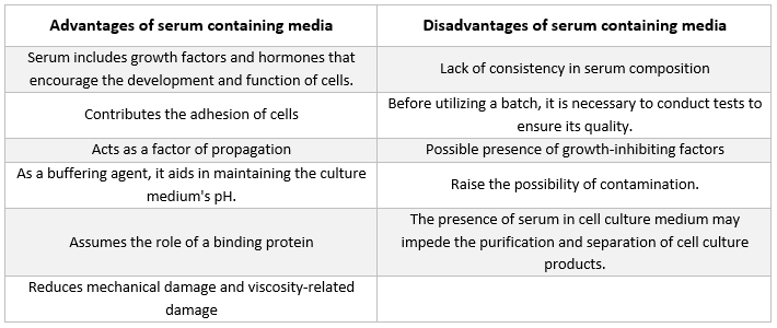
Môi trường chứa huyết thanh

Loại chất bổ sung thường gặp nhất trong môi trường dùng để nuôi cấy tế bào động vật là huyết thanh bò thai. Chất này được thêm vào môi trường nuôi cấy như một chất bổ sung giá rẻ nhằm đạt được điều kiện phát triển tốt nhất có thể. Ngoài vai trò là chất vận chuyển hoặc chất chelat hóa cho các chất dinh dưỡng không ổn định hoặc không tan trong nước, hormone và các yếu tố tăng trưởng, chất ức chế protease và các chất khác, huyết thanh còn liên kết và trung hòa các phân tử có hại.

Môi trường không có huyết thanh

Sự hiện diện của huyết thanh trong môi trường nuôi cấy có một số nhược điểm và có khả năng gây ra những sai sót lớn trong việc giải thích kết quả nghiên cứu miễn dịch [2, 3]. Đã có nhiều loại môi trường nuôi cấy không chứa huyết thanh khác nhau được tạo ra [4, 5]. Các môi trường này thường được thiết kế đặc biệt để hỗ trợ nuôi cấy một loại tế bào cụ thể, chẳng hạn như Knockout Serum Replacement và Knockout DMEM của Thermo Fisher Scientific, cùng với môi trường mTESR của Stem Cell Technologies [6], dành cho tế bào gốc [7].

Ngoài ra, các môi trường này còn chứa một lượng xác định các yếu tố tăng trưởng tinh khiết, lipoprotein và các protein khác, vốn thường được cung cấp bởi huyết thanh [8]. Các môi trường này thường được gọi là “môi trường nuôi cấy xác định” vì các thành phần cấu thành chúng đã được hiểu rõ.

Môi trường nuôi cấy được xác định về mặt hóa học

Các môi trường này bao gồm các thành phần vô cơ và hữu cơ siêu tinh khiết, không bị nhiễm bất kỳ loại tạp chất nào. Chúng cũng có thể bao gồm các chất bổ sung protein tinh khiết, chẳng hạn như các yếu tố tăng trưởng.

 Việc biến đổi gen của vi khuẩn hoặc nấm men, cùng với việc bổ sung các axit béo, vitamin, cholesterol và axit amin cụ thể, dẫn đến việc sản xuất các thành phần của chúng [9].

Môi trường không chứa protein

Môi trường không chứa protein là những môi trường hoàn toàn không chứa protein mà chỉ bao gồm các thành phần phi protein. So với môi trường có bổ sung huyết thanh, việc sử dụng môi trường không bổ sung protein thúc đẩy sự tăng sinh tế bào và biểu hiện protein mạnh mẽ hơn, đồng thời giúp việc tinh chế bất kỳ sản phẩm nào được tạo ra trong quá trình hạ lưu trở nên dễ dàng hơn [10-12]. Protein không được bao gồm trong các công thức như MEM và RPMI-1640. Tuy nhiên, có thể bổ sung protein nếu cần thiết.

Môi trường nuôi cấy và các thành phần cơ bản của nó

Môi trường nuôi cấy thương mại có thể được mua dưới dạng bột hoặc chất lỏng và thường chứa nhiều chất dinh dưỡng khác nhau như axit amin, glucose, muối, vitamin và các chất bổ sung dinh dưỡng khác. 

Nhu cầu về các thành phần này khác nhau đối với từng dòng tế bào, và những sự khác biệt này là nguyên nhân dẫn đến sự đa dạng về công thức môi trường nuôi cấy. Mỗi thành phần đảm nhiệm một chức năng nhất định, sẽ được trình bày trong các đoạn sau:

Hệ thống đệm

Để duy trì điều kiện sinh trưởng tối ưu, pH phải được kiểm soát, thường được thực hiện bằng một trong hai hệ thống đệm sau:

Hệ thống đệm tự nhiên

Tỷ lệ CO2/H2CO3 trong khí quyển bằng với tỷ lệ trong môi trường nuôi cấy, tạo ra một cơ chế đệm tự nhiên. Để duy trì cơ chế đệm tự nhiên này, các mẫu nuôi cấy phải được giữ trong môi trường không khí có nồng độ CO2 từ 5-10%, điều này thường được thực hiện bằng cách sử dụng tủ ấp CO2. Một trong những ưu điểm lớn nhất của việc sử dụng chất đệm tự nhiên là chi phí rẻ và an toàn.

HEPES

Đệm hóa học sử dụng ion lưỡng tính HEPES có khả năng đệm cao hơn trong phạm vi pH 7,2-7,4 và không cần môi trường khí được điều chỉnh. Đối với một số loại tế bào cụ thể, liều lượng HEPES cao hơn có thể gây hại. Môi trường nuôi cấy có chứa HEPES cũng dễ bị ảnh hưởng bởi tác động quang độc của ánh sáng huỳnh quang hơn nhiều [13].

Phenol Red

Chất chỉ thị pH phenol red thường được bổ sung vào môi trường nuôi cấy thương mại, cho phép theo dõi liên tục giá trị pH. Khi tế bào phát triển, các chất chuyển hóa do chúng sản sinh ra gây ra sự thay đổi pH và do đó làm thay đổi màu sắc của môi trường nuôi cấy. Phenol Red có tác động kép lên màu sắc của môi trường nuôi cấy: chuyển sang màu vàng ở pH axit và màu tím ở pH kiềm. pH 7,4 – giá trị tối ưu cho nuôi cấy tế bào – khiến môi trường nuôi cấy có màu đỏ huỳnh quang.

Tuy nhiên, phenol red có một số nhược điểm: Thứ nhất, phenol red có khả năng mô phỏng hoạt động của một số hormone steroid, chủ yếu là estrogen [14]. Do đó, khi nghiên cứu các tế bào nhạy cảm với estrogen như mô vú, nên sử dụng môi trường nuôi cấy không chứa phenol red. Sự cân bằng natri-kali bị phá vỡ do sự hiện diện của phenol red trong một số công thức không có huyết thanh. Việc thêm huyết thanh hoặc hormone tuyến yên bò vào môi trường nuôi cấy có thể khắc phục tác động này [15]. Thứ ba, việc phát hiện trong các thí nghiệm đo lưu lượng tế bào bị cản trở do sự hiện diện của phenol red.

Muối vô cơ

Môi trường nuôi cấy chứa muối vô cơ, chẳng hạn như ion natri, kali và canxi, giúp duy trì cân bằng thẩm thấu và điều chỉnh điện thế màng tế bào.

Axit amin

Vì axit amin là thành phần cơ bản của protein, chúng là thành phần thiết yếu của mọi môi trường nuôi cấy tế bào từng được phát minh. Do tế bào không thể tự sản xuất một số axit amin nhất định, nên điều quan trọng là môi trường nuôi cấy phải chứa các axit amin thiết yếu. Chúng cần thiết cho sự nhân lên của tế bào, và nồng độ của chúng quyết định mật độ tế bào tối đa có thể đạt được. Đặc biệt, L-glutamine, một axit amin thiết yếu, có vai trò đặc biệt quan trọng.

L-glutamine đóng vai trò là nguồn năng lượng thứ cấp cho quá trình chuyển hóa và cung cấp nitơ cho việc sản xuất NAD, NADPH và các nucleotide. Do L-glutamine là một axit amin không ổn định, theo thời gian sẽ chuyển hóa thành dạng mà tế bào không thể sử dụng được, nên nó phải được bổ sung vào môi trường nuôi cấy.

Ngoài ra, các axit amin không thiết yếu có thể được bổ sung vào môi trường nuôi cấy để bù đắp lượng đã bị tiêu hao trong quá trình sinh trưởng. Sự sinh trưởng của tế bào được thúc đẩy và khả năng sống sót của chúng được tăng cường khi môi trường nuôi cấy được bổ sung các axit amin không thiết yếu.

Carbohydrate

Carbohydrate dưới dạng đường là nguồn năng lượng chính. Ngoài các loại đường phổ biến như glucose và galactose, nhiều môi trường nuôi cấy còn chứa maltose và fructose.

Protein và peptit

Albumin, transferrin và fibronectin là các protein và peptit được sử dụng phổ biến nhất. Chúng đặc biệt quan trọng trong các môi trường nuôi cấy không chứa huyết thanh. Albumin, transferrin, aprotinin, fetuin và fibronectin là một số protein có thể tìm thấy trong huyết thanh, vốn là nguồn cung cấp protein dồi dào.

Albumin là protein chính có trong máu, có chức năng liên kết và vận chuyển các chất khác nhau, bao gồm nước, muối, axit béo tự do, hormone và vitamin, giữa các cơ quan và tế bào khác nhau. Khả năng liên kết với các chất hóa học của albumin khiến nó trở thành một ứng cử viên hiệu quả để loại bỏ các hợp chất có hại khỏi môi trường nuôi cấy tế bào.

Aprotinin là chất bảo vệ trong hệ thống nuôi cấy tế bào, do nó ổn định ở pH trung tính và axit, cũng như chịu được nhiệt độ cao và sự phân hủy có thể do các enzym proteolytic gây ra. Chất này có khả năng ức chế một số protease serine, bao gồm trypsin và các loại khác.

Fetuin là một glycoprotein có thể được phát hiện với lượng cao hơn trong huyết thanh của thai nhi và động vật sơ sinh so với huyết thanh của động vật trưởng thành. Ngoài ra, nó còn hoạt động như một chất ức chế protease serine. Protein fibronectin là thành phần thiết yếu trong quá trình bám dính tế bào. Transferrin là một loại protein vận chuyển sắt và chịu trách nhiệm cung cấp sắt cho màng tế bào.

Axit béo và lipid

Chúng đóng vai trò quan trọng trong môi trường nuôi cấy không có huyết thanh khi huyết thanh vắng mặt.

Vitamin

Nhiều loại vitamin là cần thiết cho sự phát triển và sinh sôi của tế bào. Tế bào không thể tự sản xuất vitamin với lượng đủ nên vitamin là chất bổ sung dinh dưỡng thiết yếu trong nuôi cấy mô.

Trong nuôi cấy tế bào, huyết thanh là nguồn vitamin chính; tuy nhiên, môi trường nuôi cấy cũng được bổ sung các loại vitamin khác nhau để phù hợp với từng loại tế bào cụ thể. Thông thường, các vitamin nhóm B được sử dụng để kích thích sự tăng trưởng.

Các nguyên tố vi lượng

Các nguyên tố hóa học như đồng, kẽm, selen và các chất trung gian của axit tricarboxylic được gọi là nguyên tố vi lượng. Nguyên tố vi lượng thường được bổ sung vào môi trường nuôi cấy không chứa huyết thanh để thay thế những nguyên tố thường có trong huyết thanh. Những nguyên tố này là các thành phần hóa học quan trọng cần thiết cho sự phát triển khỏe mạnh của tế bào. Nhiều phản ứng sinh hóa phụ thuộc vào các vi chất dinh dưỡng nhất định, chẳng hạn như hoạt động của enzyme.

Chất bổ sung môi trường nuôi cấy

Môi trường nuôi cấy hoàn chỉnh được đề xuất cho một số dòng tế bào nhất định cần có các thành phần bổ sung không có trong môi trường cơ bản và huyết thanh. Các chất bổ sung này hỗ trợ sự phát triển của tế bào và chức năng trao đổi chất thích hợp.

Mặc dù hormone, yếu tố tăng trưởng và các phân tử tín hiệu là yếu tố thiết yếu cho sự nhân lên thích hợp của các dòng tế bào cụ thể, nhưng luôn cần tuân thủ các biện pháp phòng ngừa sau: Vì việc bổ sung các chất này có thể làm thay đổi độ thẩm thấu của môi trường nuôi cấy hoàn chỉnh, từ đó có thể ức chế sự phát triển của tế bào, nên luôn cần kiểm tra lại độ thẩm thấu sau khi bổ sung. Đối với phần lớn các dòng tế bào, độ thẩm thấu tối ưu dao động trong khoảng từ 260 đến 320 mOSM/kg.

Kháng sinh

Kháng sinh thường được sử dụng để ức chế sự phát triển của các tác nhân gây ô nhiễm vi khuẩn và nấm [16], mặc dù chúng không phải là yếu tố thiết yếu cho sự phát triển của tế bào. Do kháng sinh có thể che giấu sự nhiễm bẩn bởi mycoplasma và vi khuẩn kháng thuốc, việc sử dụng thường xuyên kháng sinh không được khuyến nghị trong nuôi cấy tế bào [17, 18].

Ngoài ra, kháng sinh có thể làm rối loạn quá trình trao đổi chất của các tế bào quá mẫn cảm. Các hỗn hợp penicillin-streptomycin do MilliporeSigma và Life Technologies sản xuất thường được sử dụng. Plasmocin đã được sử dụng trong nuôi cấy các dòng tế bào u thần kinh đệm TS603, TS516 và BT260 [19], và đã được chứng minh là có hiệu quả trong việc loại bỏ nhiễm mycoplasma (20).

Huyết thanh

Albumin, các yếu tố tăng trưởng và các chất ức chế tăng trưởng đều có trong huyết thanh. Huyết thanh là một trong những thành phần quan trọng nhất của môi trường nuôi cấy tế bào vì nó cung cấp axit amin, protein, vitamin (đặc biệt là các vitamin tan trong chất béo như A, D, E và K), carbohydrate, lipid, hormone, các yếu tố tăng trưởng, khoáng chất và các nguyên tố vi lượng.

Huyết thanh từ nguồn bò thai và bê thường được sử dụng để thúc đẩy sự phát triển của tế bào nuôi cấy. Huyết thanh bò thai là nguồn cung cấp dồi dào các yếu tố tăng trưởng và phù hợp cho việc nhân bản tế bào cũng như phát triển các tế bào nhạy cảm. Do khả năng thúc đẩy tăng trưởng bị suy giảm, huyết thanh bê được sử dụng trong các thí nghiệm ức chế tiếp xúc. Môi trường nuôi cấy thông thường thường chứa từ 2% đến 10% huyết thanh. Việc bổ sung huyết thanh vào môi trường nuôi cấy phục vụ các mục đích sau [21]:

  • Huyết thanh cung cấp các chất dinh dưỡng thiết yếu cho tế bào (cả ở dạng dung dịch và gắn với protein).

  • Huyết thanh chứa một số yếu tố tăng trưởng và hormone tham gia vào quá trình thúc đẩy tăng trưởng và hoạt động chuyên biệt của tế bào.

  • Nó cung cấp nhiều protein liên kết, như albumin và transferrin, giúp vận chuyển các chất hóa học khác vào tế bào. Ví dụ, albumin vận chuyển chất béo, vitamin, hormone, v.v. vào tế bào.

  • Nó cũng cung cấp các protein, chẳng hạn như fibronectin, giúp tăng khả năng bám dính của tế bào vào chất nền. Ngoài ra, nó còn tạo ra các yếu tố lan tỏa giúp tế bào mở rộng trước khi phân chia.

  • Nó cung cấp các chất ức chế protease giúp ngăn chặn quá trình phân giải protein trong tế bào.

  • Nó cũng chứa các khoáng chất như Na+, K+, Zn2+ và Fe2+.

  • Nó làm tăng độ nhớt của môi trường nuôi cấy, từ đó bảo vệ tế bào khỏi tổn thương cơ học trong quá trình khuấy trộn khi nuôi cấy tế bào trong dung dịch lơ lửng.

  • Nó cũng đóng vai trò như một chất đệm.
Tài liệu tham khảo

[1] Morgan J, Morton H, Parker R. Dinh dưỡng của tế bào động vật trong nuôi cấy mô; các nghiên cứu ban đầu về môi trường nuôi cấy tổng hợp. Proc Soc Exp Biol Med. 1950;73:1-8

[2] Kerbel R, Blakeslee D. Sự hấp phụ nhanh chóng của một thành phần huyết thanh bò thai bởi các tế bào động vật có vú trong nuôi cấy. Một nguồn tiềm ẩn gây ra hiện tượng giả trong các nghiên cứu về huyết thanh kháng thể đối với các kháng nguyên đặc hiệu tế bào. Immunology. 1976;31:881-91

[3] Sula K, Draber P, Nouza K. Việc bổ sung huyết thanh vào môi trường dùng để chuẩn bị huyền phù tế bào là một nguồn có thể gây ra sai lệch trong các phản ứng miễn dịch tế bào được nghiên cứu thông qua thử nghiệm hạch bạch huyết vùng gối. Tạp chí Di truyền Miễn dịch. 1980;7:483-9

[4] Mariani E, Mariani A, Monaco M, Lalli E, Vitale M, Facchini A. Môi trường nuôi cấy không chứa huyết thanh thương mại: sự phát triển của tế bào lai và sản xuất kháng thể đơn dòng. Tạp chí Phương pháp Miễn dịch học. 1991;145:175-83

[5] Barnes D, Sato G. Các phương pháp nuôi cấy tế bào trong môi trường không chứa huyết thanh. Anal Biochem. 1980;102:255-70

[6] Yu H, Lu S, Gasior K, Singh D, Vazquez Sanchez S, Tapia O, và cộng sự. Các protein chaperone HSP70 vận chuyển TDP-43 không chứa RNA vào các vỏ cầu lỏng nội nhân có tính dị hướng. Science. 2021;371:

[7] Meharena H, Marco A, Dileep V, Lockshin E, Akatsu G, Mullahoo J, và cộng sự. Quá trình lão hóa do hội chứng Down gây ra làm rối loạn cấu trúc nhân của các tế bào tiền thân thần kinh. Cell Stem Cell. 2022;29:116-130.e7

[8] Iscove N, Melchers F. Thay thế hoàn toàn huyết thanh bằng albumin, transferrin và lipid đậu nành trong môi trường nuôi cấy các tế bào lympho B phản ứng với lipopolysaccharide. J Exp Med. 1978;147:923-33

[9] Stoll T, Muhlethaler K, von Stockar U, Marison I. Cải tiến có hệ thống môi trường nuôi cấy không chứa protein và được định nghĩa hóa học dành cho sự phát triển của tế bào hybridoma và sản xuất kháng thể đơn dòng. J Biotechnol. 1996;45:111-23

[10] Darfler F. Môi trường không chứa protein để nuôi cấy tế bào lai và các tế bào khác của hệ miễn dịch. In Vitro Cell Dev Biol. 1990;26:769-78

[11] Barnes D, Sato G. Nuôi cấy tế bào không huyết thanh: một phương pháp thống nhất. Cell. 1980;22:649-55

[12] Hamilton W, Ham R. Sự phát triển dòng tế bào chuột hamster Trung Quốc trong môi trường không chứa protein. In Vitro. 1977;13:537-47

[13] Zigler J, Lepe Zuniga J, Vistica B, Gery I. Phân tích tác dụng độc tế bào của môi trường nuôi cấy chứa HEPES khi tiếp xúc với ánh sáng. In Vitro Cell Dev Biol. 1985;21:282-7

[14] Berthois Y, Katzenellenbogen J, Katzenellenbogen B. Chất Phenol red trong môi trường nuôi cấy mô là một estrogen yếu: những hàm ý liên quan đến việc nghiên cứu các tế bào nhạy cảm với estrogen trong nuôi cấy. Proc Natl Acad Sci U S A. 1986;83:2496-500

[15] Karmiol S. Phát triển môi trường nuôi cấy không có huyết thanh. Trong: Master JRW, biên tập. Animal Cell culture, ấn bản thứ 3. Oxford: Oxford University Press; 2000.

[16] Perlman D. Việc sử dụng kháng sinh trong môi trường nuôi cấy tế bào. Methods Enzymol. 1979;58:110-6

[17] McGarrity G. Sự lây lan và kiểm soát nhiễm trùng mycoplasma trong nuôi cấy tế bào. In Vitro. 1976;12:643-8

[18] Masters J, Stacey G. Thay đổi môi trường nuôi cấy và nhân giống dòng tế bào. Nat Protoc. 2007;2:2276-84

[19] Chakraborty A, Laukka T, Myllykoski M, Ringel A, Booker M, Tolstorukov M, và cộng sự. Enzym demethylase histone KDM6A trực tiếp cảm nhận oxy để điều chỉnh cấu trúc chromatin và số phận tế bào. Science. 2019;363:1217-1222

[20] Molla Kazemiha V, Azari S, Amanzadeh A, Bonakdar S, Shojaei Moghadam M, Habibi Anbouhi M, và cộng sự. Hiệu quả của Plasmocin™ trên các dòng tế bào động vật có vú bị nhiễm mollicutes so với các loại kháng sinh thường được sử dụng trong nuôi cấy tế bào: kinh nghiệm tại địa phương. Cytotechnology. 2011;63:609-20

[21] Kragh Hansen U. Các khía cạnh phân tử của sự liên kết ligand với albumin huyết thanh. Pharmacol Rev. 1981;33:17-53