
Skip to content

Phương tiện truyền thông & Dung dịch phản ứng

40 products

Accutase Cell Detachment Solution - 100 ml

Accutase Cell Detachment Solution with EDTA and Phenol Red – 100 ml Accutase is a ready-to-use, sterile-filtered cell detachment solution designed as a gentle alternative to trypsin/EDTA for dissociating adherent mammalian cells from standard tissue culture plasticware and adhesion-coated surfaces. It combines proteolytic and collagenolytic enzyme activity in a balanced salt solution to deliver effective yet controlled dissociation, preserving cell-surface proteins and supporting high post-passage viability and rapid reattachment. The Accutase formulation is based on Dulbecco’s phosphate-buffered saline (DPBS) with EDTA and phenol red as a visual pH indicator. The enzymes are of non-mammalian and non-bacterial origin, making Accutase particularly well suited to stem cell research, vaccine workflows, and any application where animal- or microbially-derived contaminants must be minimised. The solution auto-inhibits at 37 °C, so no neutralising reagent or serum-containing medium is required after detachment – cells can be transferred directly into fresh medium. Key Features • Ready-to-use 1x sterile-filtered liquid – no dilution or reconstitution required • Combined proteolytic and collagenolytic enzyme activity for gentle dissociation • Each batch standardised to a defined dissociation activity for lot-to-lot consistency • Non-mammalian and non-bacterial enzyme origin • Auto-inhibits at 37 °C – no neutralising solution needed • Formulated in Dulbecco’s PBS with EDTA • Phenol red included as visual pH indicator • pH 6.8 – 7.8 Typical Applications Accutase gently dissociates a wide variety of adherent and sensitive cell types, including human embryonic stem cells (hESCs), human induced pluripotent stem cells (iPSCs), neural stem cells, primary neurons, and routinely cultured adherent lines such as HeLa, HEK 293, CHO, MDCK, Vero, NIH/3T3, BHK-21 and A549. Typical use cases include: • Routine subculture and passaging of adherent mammalian cells • Gentle single-cell dissociation of hESCs, iPSCs and other sensitive lines • Sample preparation for flow cytometry and FACS analysis • Analysis of cell-surface markers where epitope integrity matters • Cell migration, proliferation and apoptosis assays • Quiescence assays by serum starvation and oncogene transfection studies • Tumor cell and neural crest cell migration assays • Production scale-up in bioreactor workflows For routine work, apply approximately 10 ml of Accutase per 75 cm2 of culture surface and incubate for 5–10 minutes at room temperature. The optimal incubation time should be determined for each cell line and should not exceed one hour. Prior to addition, rinse the cell layer with a Ca2+/Mg2+-free salt solution such as DPBS without calcium and magnesium to remove residual serum and divalent cations. Handling & Storage Store the unopened bottle frozen at -15 °C or below. Thaw either at room temperature or overnight at +2 °C to +8 °C. Do not thaw Accutase in a 37 °C water bath, as elevated temperatures reduce enzyme activity. After thawing, the solution can be stored for up to 2 months at +2 °C to +8 °C; do not store at room temperature. Do not pre-warm the reagent to 37 °C before application – add it directly to washed cells at room temperature. For long-term shelf life, single-use aliquoting is recommended to avoid repeated thaw cycles. Always work under aseptic conditions. Quality Manufactured under strict quality standards. Each batch of Accutase is sterile-filtered and tested for sterility, pH, appearance and dissociation activity to ensure consistent, reproducible performance from lot to lot. Product Specifications Specification Detail Product typeCell detachment / dissociation reagent FormatSterile-filtered liquid, ready-to-use Volume100 ml Working concentration1x (ready-to-use) Enzyme activityCombined proteolytic and collagenolytic Enzyme originNon-mammalian and non-bacterial Buffer systemDulbecco’s PBS with EDTA pH indicatorPhenol red pH range6.8 – 7.8 AppearanceClear, pale red to orange solution Storage temperature-15 °C or below Stability after thawingUp to 2 months at +2 °C to +8 °C Recommended use volume~10 ml per 75 cm2 culture surface Typical incubation time5 – 10 minutes at room temperature Shipping conditionsFrozen on dry ice Intended useFor research use and further manufacturing only Formulation (Composition per Liter) Accutase is a registered trademark of Innovative Cell Technologies, Inc.

€75.00

excl. tax and shipping

Accutase Cell Detachment Solution - 500 ml

Accutase Cell Detachment Solution with EDTA and Phenol Red – 100 ml Accutase is a ready-to-use, sterile-filtered cell detachment solution designed as a gentle alternative to trypsin/EDTA for dissociating adherent mammalian cells from standard tissue culture plasticware and adhesion-coated surfaces. It combines proteolytic and collagenolytic enzyme activity in a balanced salt solution to deliver effective yet controlled dissociation, preserving cell-surface proteins and supporting high post-passage viability and rapid reattachment. The Accutase formulation is based on Dulbecco’s phosphate-buffered saline (DPBS) with EDTA and phenol red as a visual pH indicator. The enzymes are of non-mammalian and non-bacterial origin, making Accutase particularly well suited to stem cell research, vaccine workflows, and any application where animal- or microbially-derived contaminants must be minimised. The solution auto-inhibits at 37 °C, so no neutralising reagent or serum-containing medium is required after detachment – cells can be transferred directly into fresh medium. Key Features • Ready-to-use 1x sterile-filtered liquid – no dilution or reconstitution required • Combined proteolytic and collagenolytic enzyme activity for gentle dissociation • Each batch standardised to a defined dissociation activity for lot-to-lot consistency • Non-mammalian and non-bacterial enzyme origin • Auto-inhibits at 37 °C – no neutralising solution needed • Formulated in Dulbecco’s PBS with EDTA • Phenol red included as visual pH indicator • pH 6.8 – 7.8 Typical Applications Accutase gently dissociates a wide variety of adherent and sensitive cell types, including human embryonic stem cells (hESCs), human induced pluripotent stem cells (iPSCs), neural stem cells, primary neurons, and routinely cultured adherent lines such as HeLa, HEK 293, CHO, MDCK, Vero, NIH/3T3, BHK-21 and A549. Typical use cases include: • Routine subculture and passaging of adherent mammalian cells • Gentle single-cell dissociation of hESCs, iPSCs and other sensitive lines • Sample preparation for flow cytometry and FACS analysis • Analysis of cell-surface markers where epitope integrity matters • Cell migration, proliferation and apoptosis assays • Quiescence assays by serum starvation and oncogene transfection studies • Tumor cell and neural crest cell migration assays • Production scale-up in bioreactor workflows For routine work, apply approximately 10 ml of Accutase per 75 cm2 of culture surface and incubate for 5–10 minutes at room temperature. The optimal incubation time should be determined for each cell line and should not exceed one hour. Prior to addition, rinse the cell layer with a Ca2+/Mg2+-free salt solution such as DPBS without calcium and magnesium to remove residual serum and divalent cations. Handling & Storage Store the unopened bottle frozen at -15 °C or below. Thaw either at room temperature or overnight at +2 °C to +8 °C. Do not thaw Accutase in a 37 °C water bath, as elevated temperatures reduce enzyme activity. After thawing, the solution can be stored for up to 2 months at +2 °C to +8 °C; do not store at room temperature. Do not pre-warm the reagent to 37 °C before application – add it directly to washed cells at room temperature. For long-term shelf life, single-use aliquoting is recommended to avoid repeated thaw cycles. Always work under aseptic conditions. Quality Manufactured under strict quality standards. Each batch of Accutase is sterile-filtered and tested for sterility, pH, appearance and dissociation activity to ensure consistent, reproducible performance from lot to lot. Product Specifications Specification Detail Product typeCell detachment / dissociation reagent FormatSterile-filtered liquid, ready-to-use Volume100 ml Working concentration1x (ready-to-use) Enzyme activityCombined proteolytic and collagenolytic Enzyme originNon-mammalian and non-bacterial Buffer systemDulbecco’s PBS with EDTA pH indicatorPhenol red pH range6.8 – 7.8 AppearanceClear, pale red to orange solution Storage temperature-15 °C or below Stability after thawingUp to 2 months at +2 °C to +8 °C Recommended use volume~10 ml per 75 cm2 culture surface Typical incubation time5 – 10 minutes at room temperature Shipping conditionsFrozen on dry ice Intended useFor research use and further manufacturing only Formulation (Composition per Liter) Accutase is a registered trademark of Innovative Cell Technologies, Inc.

€310.00

excl. tax and shipping

Antibiotic/Antimycotic Solution (100x)

Volume: 100 ml Storage: ≤-15°C Sterility: Sterile-filtered Antibiotic/Antimycotic Solution (100x) is a sterile, ready-to-use concentrate designed to reduce microbial contamination risks in cell culture and related laboratory applications. This 100x solution contains a well-established combination of penicillin, streptomycin, and amphotericin B—providing broad-spectrum antimicrobial activity against Gram-positive and Gram-negative bacteria, yeasts, and filamentous fungi. The formulation is suitable for use in eukaryotic cell cultures, bacterial media, and other contamination-sensitive systems, supporting clean and consistent lab operations. Application and Benefits Optimized for routine research protocols, this solution is widely used to maintain aseptic conditions in cell culture workflows. It offers reliable performance in contamination-sensitive environments, helping researchers reduce the risk of microbial overgrowth without compromising cell health or experimental reproducibility. The sterile-filtered formulation eliminates the need for additional solubilization steps, supporting streamlined media preparation and reducing variability in daily lab procedures. Usage and Compatibility To achieve standard working concentrations, dilute the solution 1:100 into your complete culture medium. The product is compatible with a broad range of mammalian cell lines and basal media. With consistent stock availability, researchers benefit from dependable supply continuity and simplified logistics planning. The solution should be stored at ≤ –15 °C and protected from repeated freeze-thaw cycles to maintain stability. For research use only. Not for use in diagnostic or therapeutic procedures. Not for use in humans or animals.

€45.00

excl. tax and shipping

DMEM:Ham's F12 (1:1), w: 3.1 g/L Glucose, w: 2.5 mM L-Glutamine, w: 15 mM HEPES, w: 0.5 mM Sodium pyruvate, w: 1.2 g/L NaHCO3

DMEM:Ham's F12 is a widely recognized and extensively utilized basal medium in cell culture for biological research. It serves as a fundamental source of nutrients for the growth of various mammalian cell lines, particularly when supplemented with Fetal Bovine Serum (FBS). This unique formulation combines Dulbecco's Modified Eagle Medium (DMEM) and Ham's F-12 (Ham's Nutrient Mixture F-12) in a precise 1:1 ratio. The addition of L-glutamine further enhances its composition. DMEM, derived from Eagle's Minimal Essential Medium (EMEM), offers an increased concentration of amino acids and vitamins compared to its predecessor. In contrast, Ham's F-12 is based on Ham's F-10 medium, providing a complementary set of essential components. To support optimal cell growth, it is common practice to supplement DMEM:Ham's F12 with FBS at a typical concentration of 5-10%. This addition is necessary as the medium lacks growth hormones, lipids, and proteins crucial for cellular development. DMEM:Ham's F12 incorporates a pH buffer system and is often supplemented with phenol red, a pH indicator. Cultured cells in DMEM:Ham's F12, or any medium utilizing the bicarbonate buffer system, require a controlled CO2 environment of 5-10% to maintain appropriate pH levels. Quality Control • Sterile-filtered Storage and Shelf Life • Store at +2°C to +8°C, protected from light. • Once opened, store at 4°C and use within 6–8 weeks. Shipping Conditions • Ambient temperature Maintenance • Keep refrigerated at +2°C to +8°C in the dark. Avoid freezing and frequent warming to +37°C, as it reduces product quality. • Do not heat the medium beyond 37°C or use uncontrolled heat sources such as microwave appliances. • If only part of the medium is to be used, remove the required amount and warm it to room temperature before use. Composition Category Components Concentration (mg/L) Amino Acids Glycine 18.75 L-Alanine 4.45 L-Arginine HCl 147.50 L-Asparagine H₂O 7.50 L-Aspartic Acid 6.65 L-Cysteine HCl H₂O 17.56 L-Cystine 2 HCl 31.29 L-Glutamic Acid 7.35 L-Glutamine 365.00 L-Histidine HCl H₂O 31.48 L-Isoleucine 54.47 L-Leucine 59.05 L-Lysine HCl 91.25 L-Methionine 17.24 L-Phenylalanine 35.48 L-Proline 17.25 L-Serine 26.25 L-Threonine 53.45 L-Tryptophan 9.02 L-Tyrosine 2 Na 2 H2O 55.79 L-Valine 52.85 Vitamins D-Biotin 0.0035 Choline Chloride 8.98 D-Calcium Pantothenate 2.24 Folic Acid 2.66 myo-Inositol 12.60 Nicotinamide 2.02 Pyridoxine HCl 0.031 Pyridoxal HCl 2.00 Riboflavin 0.219 Thiamine HCl 2.17 Vitamin B12 0.68 Inorganic Salts CaCl2 2 H2O 154.50 CuSO4 5 H2O 0.0013 Fe(NO3)3 9 H2O 0.05 FeSO4 7 H2O 0.417 KCl 311.80 MgCl2 6 H2O 61.20 MgSO4 7 H2O 100.00 NaCl 6996.00 NaHCO3 1200.00 Na2HPO4 71.02 NaH2PO4 2 H2O 70.87 ZnSO4 7 H2O 0.432 Other Components D-Glucose 3151.00 Hypoxanthine 2.40 HEPES 3574.50 Linoleic Acid 0.042 Lipoic Acid 0.105 Phenol Red Sodium Salt 8.63 Putrescine 2 HCl 0.081 Sodium Pyruvate 55.00 Thymidine 0.365

€25.00

excl. tax and shipping

DMEM, w: 4.5 g/L Glucose, w: 4 mM L-Glutamine, w: 3.7 g/L NaHCO3, w: 1.0 mM Sodium pyruvate

DMEM (Dulbecco's Modified Eagle Medium) is a highly versatile and widely utilized basal medium designed to support the growth of a diverse range of mammalian cells in biological research. It serves as an ideal medium for culturing primary fibroblasts, neurons, glial cells, HUVECs, smooth muscle cells, as well as popular cell lines like HeLa, 293, Cos-7, and PC-12. What sets DMEM apart from other media is its unique composition. It contains an impressive fourfold increase in amino acid and vitamin concentration compared to the original Eagle's Minimal Essential Medium. Initially developed with low glucose (1 g/L) and sodium pyruvate, DMEM is frequently employed with higher glucose levels, either with or without sodium pyruvate. Notably, DMEM does not contain proteins, lipids, or growth factors, necessitating supplementation. To achieve optimal growth, a common approach is to supplement DMEM with 10% Fetal Bovine Serum (FBS). Additionally, DMEM employs a sodium bicarbonate buffer system, requiring a 5-10% CO2 environment to maintain a physiological pH. Dulbecco's Modified Eagle Medium (DMEM) is highly regarded among defined media for cell and tissue culture, catering to the growth needs of various adherent cell phenotypes. It surpasses the original Eagle's Medium, developed in the 1950s for cultivating chicken cells, through the enhanced supplementary formulation known as Dulbecco's modification. This modification significantly elevates the content of select amino acids and vitamins up to fourfold compared to the original medium. In the field of cell culture, DMEM plays a vital role as a growth medium for different cell types, including primary cells, stem cells, and transformed cells. Researchers also employ the modified version of DMEM for a wide array of research applications, such as drug discovery, tissue engineering, and the study of cell signaling pathways. Quality Control • Sterile-filtered Storage and Shelf Life • Store at +2°C to +8°C, protected from light. • Once opened, store at 4°C and use within 6–8 weeks. Shipping Conditions • Ambient temperature Maintenance • Keep refrigerated at +2°C to +8°C in the dark. Avoid freezing and frequent warming to +37°C, as it reduces product quality. • Do not heat the medium beyond 37°C or use uncontrolled heat sources such as microwave appliances. • If only part of the medium is to be used, remove the required amount and warm it to room temperature before use. Composition Category Components Concentration (mg/L) Amino Acids Glycine 30.00 L-Arginine HCl 84.00 L-Cystine 2 HCl 62.57 L-Glutamine 584.00 L-Histidine HCl H2O 42.00 L-Isoleucine 105.00 L-Leucine 105.00 L-Lysine HCl 146.00 L-Methionine 30.00 L-Phenylalanine 66.00 L-Serine 42.00 L-Threonine 95.00 L-Tryptophan 16.00 L-Tyrosine 2 Na 2H2O 103.79 L-Valine 94.00 Vitamins Choline chloride 4.00 D-Calcium Pantothenate 4.00 Folic Acid 4.00 myo-Inositol 7.20 Nicotinamide 4.00 Pyridoxal HCl 4.00 Riboflavin 0.40 Thiamine HCl 4.00 Inorganic Salts CaCl2 2 H2O 265.00 Fe(NO3)3 9 H2O 0.10 KCl 400.00 MgSO4 7H2O 200.10 NaCl 6400.00 NaHCO3 3700.00 NaH2PO4 2H2O 141.73 Other Components D-Glucose 4500.00 Phenol Red Sodium Salt 15.90 Sodium Pyruvate 110.00

€25.00

excl. tax and shipping

EMEM (MEM Eagle), w: 2 mM L-Glutamine, w: 2.2 g/L NaHCO3, w: EBSS

Eagle’s Minimum Essential Medium (EMEM) is one of the most widely used basal media for the cultivation of a broad range of mammalian cells, particularly adherent cell lines. Originally developed by Harry Eagle, this classic formulation contains the essential amino acids, vitamins, and inorganic salts required to support the growth of both primary cells and established cell lines under standard culture conditions. This ready-to-use, sterile-filtered liquid formulation is supplemented with Earle’s Balanced Salt Solution (EBSS), 2 mM L-glutamine, D-glucose (1.0 g/L), and 2.2 g/L sodium bicarbonate (NaHCO3), making it suitable for use in a CO2-controlled incubator atmosphere (typically 5 % CO2). The included phenol red acts as a pH indicator, allowing convenient visual monitoring of medium condition during cell culture. Key Features • Classic Eagle’s MEM formulation with Earle’s Balanced Salt Solution (EBSS) • 2 mM L-glutamine included – ready for immediate use • 2.2 g/L sodium bicarbonate – buffered for 5 % CO2 incubation • With D-glucose (1.0 g/L) as primary carbon source • With phenol red as pH indicator • Without HEPES and without sodium pyruvate • Sterile-filtered liquid medium, ready to use • pH 7.0 – 7.6 Typical Applications EMEM supports the cultivation of a wide variety of mammalian cell lines, including HeLa, HEK 293, Vero, MRC-5, L-929, BHK-21, and many primary cells. Common applications include: • Routine maintenance and expansion of adherent cell lines • Virus propagation and vaccine production workflows • Cytotoxicity and bioassay applications • Transfection and protein expression studies • Foundational research in cell biology and molecular biology For optimal cell growth, EMEM is typically supplemented with 5–10 % fetal bovine serum (FBS) and, depending on the cell line, with non-essential amino acids (NEAA) and antibiotics such as penicillin/streptomycin. Handling & Storage Store the unopened bottle at +2 °C to +8 °C, protected from light. After opening, use under aseptic conditions. L-Glutamine in solution is subject to gradual degradation – we recommend using the medium within 4 weeks of opening for best performance, or supplementing with fresh L-glutamine prior to use if stored for longer periods. Allow the medium to warm to 37 °C before adding it to cells. Quality Manufactured under strict quality standards. Each batch is tested for sterility, pH, osmolality, and endotoxin levels to ensure consistent performance in cell culture applications. Product Specifications Specification Detail Product typeMEM Product categoryCell culture media FormatLiquid SterileYes Size500 ml L-GlutamineWith L-glutamine (2 mM) GlucoseWith glucose (1.0 g/L) Sodium bicarbonateWith NaHCO3 (2.2 g/L) HEPESWithout HEPES Sodium pyruvateWithout sodium pyruvate Phenol redWith phenol red Salt solutionEarle’s Balanced Salt Solution (EBSS) pH7.0 – 7.6 Endotoxin contentNot specified Storage+2 °C to +8 °C Formulation (Composition per Liter)

€25.00

excl. tax and shipping

Freeze Medium CM-1 - 100 ml

Cytion’s Freeze Medium CM-1 is a state-of-the-art cryopreservation medium designed to ensure the highest level of cell viability and functionality post-thaw. This versatile medium is suitable for a broad spectrum of cell types, including both human and animal cells, making it an essential tool for diverse research applications. Formulated with a meticulously balanced combination of cryoprotectants and essential nutrients, Freeze Medium CM-1 minimizes ice crystal formation and cellular stress during the freezing process, thus preserving cellular integrity. Key features of Freeze Medium CM-1 include: • Broad Compatibility: Effective for a wide range of cell types, including primary cells, stem cells, and established cell lines. • High Viability: Optimized to maximize post-thaw cell recovery and viability, ensuring reliable experimental outcomes. • Ready-to-Use: Conveniently prepared and sterilized for immediate application, reducing preparation time and risk of contamination. • Enhanced Stability: Maintains consistent performance under standard cryopreservation conditions, ensuring reproducible results. • Long Shelf Life: CM-1 is a serum-containing, ready-to-use cryopreservation medium that can be stored in the refrigerator for up to one year. Using CM-1 for Freezing Cells To use CM-1 for freezing both adherent and suspension cells, follow these steps: • For adherent cells, wash and dissociate them from the culture substrate. For suspension cells, proceed directly to the next step. • Count the cells to ensure they are at the proper concentration. • Centrifuge the cells to pellet them, then resuspend in CM-1 freeze medium. • Transfer the resuspended cells into cryovials. • Use a slow-freezing method before transferring the cells to long-term storage. Method Description Steps ❄️ Manual Freezing A step-by-step method involving gradual temperature reduction to ensure cell viability. 1️⃣ Place cells in freeze medium in a 4°C freezer for 40 minutes. 2️⃣ Transfer to a -80°C freezer for 24 hours. 3️⃣ Store cells in liquid nitrogen for long-term preservation. ❄️ Using Mr. Frosty A convenient device that allows for controlled freezing rates without electrical power. 1️⃣ Prepare cells in cryovials with freeze medium. 2️⃣ Place cryovials in Mr. Frosty container. 3️⃣ Store at -80°C for 24 hours before transferring to liquid nitrogen. ❄️ Controlled-Rate Freezer A high-precision freezer by Thermo Fisher or other manufacturers designed for controlled temperature reduction. 1️⃣ Program the device to gradually decrease the temperature. 2️⃣ Place prepared cells in the freezer. 3️⃣ After the freezing cycle, transfer cells to liquid nitrogen. • Store the cryovials at temperatures below -130°C or in liquid nitrogen for long-term preservation. Ingredients • Contains FBS, DMSO, Glucose, Salts • Buffering capacity: pH = 7.2 to 7.6 Cytion’s Freeze Medium CM-1 offers a reliable solution for cryopreservation, ensuring high cell viability and functionality post-thaw for a wide range of research applications.

€99.00

excl. tax and shipping

Freeze Medium CM-1 - 50 ml

Cytion’s Freeze Medium CM-1 is a state-of-the-art cryopreservation medium designed to ensure the highest level of cell viability and functionality post-thaw. This versatile medium is suitable for a broad spectrum of cell types, including both human and animal cells, making it an essential tool for diverse research applications. Formulated with a meticulously balanced combination of cryoprotectants and essential nutrients, Freeze Medium CM-1 minimizes ice crystal formation and cellular stress during the freezing process, thus preserving cellular integrity. Key features of Freeze Medium CM-1 include: • Broad Compatibility: Effective for a wide range of cell types, including primary cells, stem cells, and established cell lines. • High Viability: Optimized to maximize post-thaw cell recovery and viability, ensuring reliable experimental outcomes. • Ready-to-Use: Conveniently prepared and sterilized for immediate application, reducing preparation time and risk of contamination. • Enhanced Stability: Maintains consistent performance under standard cryopreservation conditions, ensuring reproducible results. • Long Shelf Life: CM-1 is a serum-containing, ready-to-use cryopreservation medium that can be stored in the refrigerator for up to one year. Using CM-1 for Freezing Cells To use CM-1 for freezing both adherent and suspension cells, follow these steps: • For adherent cells, wash and dissociate them from the culture substrate. For suspension cells, proceed directly to the next step. • Count the cells to ensure they are at the proper concentration. • Centrifuge the cells to pellet them, then resuspend in CM-1 freeze medium. • Transfer the resuspended cells into cryovials. • Use a slow-freezing method before transferring the cells to long-term storage. Method Description Steps ❄️ Manual Freezing A step-by-step method involving gradual temperature reduction to ensure cell viability. 1️⃣ Place cells in freeze medium in a 4°C freezer for 40 minutes. 2️⃣ Transfer to a -80°C freezer for 24 hours. 3️⃣ Store cells in liquid nitrogen for long-term preservation. ❄️ Using Mr. Frosty A convenient device that allows for controlled freezing rates without electrical power. 1️⃣ Prepare cells in cryovials with freeze medium. 2️⃣ Place cryovials in Mr. Frosty container. 3️⃣ Store at -80°C for 24 hours before transferring to liquid nitrogen. ❄️ Controlled-Rate Freezer A high-precision freezer by Thermo Fisher or other manufacturers designed for controlled temperature reduction. 1️⃣ Program the device to gradually decrease the temperature. 2️⃣ Place prepared cells in the freezer. 3️⃣ After the freezing cycle, transfer cells to liquid nitrogen. • Store the cryovials at temperatures below -130°C or in liquid nitrogen for long-term preservation. Ingredients • Contains FBS, DMSO, Glucose, Salts • Buffering capacity: pH = 7.2 to 7.6 Cytion’s Freeze Medium CM-1 offers a reliable solution for cryopreservation, ensuring high cell viability and functionality post-thaw for a wide range of research applications.

€59.00

excl. tax and shipping

Ham's F12K Medium, w: 2.0 mM L-Glutamine, w: 2.0 mM Sodium pyruvate, w: 2.5 g/L NaHCO3

Introducing Ham's F-12K (Kaighn's) Medium, a specialized modification of Ham's F-12 medium designed to meet the unique requirements of biological research. This advanced medium offers distinct advantages, enhancing the cultivation of primary human hepatocytes, as well as rat and chicken liver cells, particularly in reduced serum conditions. Ham's F-12K (Kaighn's) Medium is carefully formulated to optimize cell culture conditions. It features an enriched composition, providing elevated levels of essential components such as amino acids and sodium pyruvate, as well as additional elements including putrescine, thymidine, hypoxanthine, and zinc. These additions enable researchers to supplement the medium with minimal serum or defined components for specific cell types, facilitating precise experimental conditions. Notably, Ham's F-12K (Kaighn's) Medium does not contain proteins or growth factors. Consequently, supplementation with growth factors and Fetal Bovine Serum (FBS) is often necessary, allowing researchers to tailor the medium to the requirements of their specific cell lines. For optimal performance, the concentration of FBS must be carefully optimized for each cell line, ensuring optimal growth and functionality. To maintain physiological pH, Ham's F-12K (Kaighn's) Medium employs a sodium bicarbonate buffer system (2.5 g/L), necessitating a controlled 5-10% CO2 environment during cultivation. This ensures the medium's pH remains within the ideal range for cell growth and viability. Quality control • pH = 7.2 +/- 0.02 at 20-25°C. • Each lot has been tested for sterility and absence of mycoplasma and bacteria. Maintenance • Keep refrigerated at +2°C to +8°C in the dark. Freezing as well as warming up to +37°C minimize the quality of the product. • Do not heat the medium to more than 37°C or use uncontrollable sources of heat (e.g., microwave appliances). • If only a part of the medium is to be used, remove this amount from the bottle and warm it up at room temperature. • Shelf life for any medium but the basic medium is 8 weeks from the date of manufacture. Composition Components mg/L Inorganic Salts Calcium chloride x 2H2O 135,24 Copper(II) sulfate x 5H2O 0,00 Iron (II) sulfate x 7H2O 0,83 Magnesium chloride x 6H2O 105,72 Magnesium sulfate x 7H2O 394,49 Potassium chloride 283,29 Potassium dihydrogen phosphate 58,52 Sodium chloride 7597,20 di-Sodium hydrogen phosphate anhydrous 115,02 Zinc sulfate x 7H2O 0,14 Other Components D(+)-Glucose anhydrous 1260,00 Hypoxanthine 4,08 DL-α-Lipoic acid 0,21 Phenol red 3,00 Putrescine x 2HCl 0,32 Sodium pyruvate 220,00 NaHCO3 2500,00 Thymidine 0,73 Amino Acids L-Alanine 17,82 L-Arginine x HCl 421,40 L-Asparagine x H2O 30,02 L-Aspartic acid 26,62 L-Cysteine x HCl x H2O 70,24 L-Glutamine 292,20 L-Glutamic acid 29,42 Glycine 15,01 L-Histidine x HCl x H2O 41,92 L-Isoleucine 7,87 L-Leucine 26,24 L-Lysine x HCl 73,04 L-Methionine 8,95 L-Phenylalanine 9,91 L-Proline 69,06 L-Serine 21,02 L-Threonine 23,82 L-Tryptophan 4,08 L-Tyrosine 10,87 L-Valine 23,42 Vitamins D(+)-Biotin 0,07 D-Calcium pantothenate 0,48 Choline chloride 13,96 Folic acid 1,32 myo-Inositol 18,02 Nicotinamide 0,04 Pyridoxine x HCl 0,06 Riboflavin 0,04 Thiamine x HCl 0,34 Vitamin B12 1,36

€108.00

excl. tax and shipping

Ham's F12 Medium, w: 1.0 mM stable Glutamine, w: 1.0 mM Sodium pyruvate, w: 1.176 g/L NaHCO3

Ham's F-12 Medium, also known as Ham's F-12 Nutrient Mix, is a widely used basal medium designed specifically for cell culture. It has been extensively employed for serum-free, single-cell plating of Chinese Hamster Ovary (CHO) cells, lung cells, and mouse L cells. Additionally, Ham’s F-12 is the medium of choice for a Clonal Toxicity Assay (CTA). One of its notable advantages is the ability to support cell growth without the need for serum supplementation. This eliminates potential interference caused by serum components, ensuring consistent and reliable experimental results. By providing a serum-free culture environment, Ham's F-12 Medium offers researchers greater control over their investigations. Another key feature of Ham's F-12 Medium is its suitability for single-cell plating. This makes it an excellent choice for a variety of cell lines, including CHO cells, lung cells, and mouse L cells. The medium's optimized nutrient composition facilitates efficient attachment and growth of individual cells, enabling the establishment of homogeneous cell cultures with improved reproducibility. Moreover, Ham's F-12 Medium has gained recognition as the preferred medium for the Clonal Toxicity Assay (CTA). This assay plays a critical role in assessing the cytotoxic effects of substances on cells. By utilizing Ham's F-12 Medium in the CTA, researchers can accurately evaluate the impact of various compounds or treatments on individual cells, providing valuable insights into toxicological profiles. Quality control • pH = 7.2 +/- 0.02 at 20-25°C. • Each lot has been tested for sterility and absence of mycoplasma and bacteria. Maintenance • Keep refrigerated at +2°C to +8°C in the dark. Freezing and warming up to +37° C minimize the quality of the product. • Do not heat the medium to more than 37° C or use uncontrollable sources of heat (e.g., microwave appliances). • If only a part of the medium is to be used, remove this amount from the bottle and warm it up at room temperature. • Shelf life for any medium except for the basic medium is 8 weeks from the date of manufacture. Composition Components mg/L Inorganic Salts Calcium chloride x 2H2O 44,00 Copper(II) sulfate x 5H2O 0,00 Iron (II) sulfate x 7H2O 0,83 Magnesium chloride x 6H2O 122,00 Potassium chloride 223,65 Sodium chloride 7599,00 di-Sodium hydrogen phosphate anhydrous 142,04 Zinc sulfate x 7H2O 0,86 Other Components D(+)-Glucose anhydrous 1801,60 Hypoxanthine 4,08 Linoleic acid 0,08 DL-α-Lipoic acid 0,21 Phenol red 1,20 Putrescine x 2HCl 0,16 Sodium pyruvate 110,00 Thymidine 0,73 NaHCO3 1176,00 Amino Acids L-Alanine 8,91 L-Arginine x HCl 210,70 L-Asparagine x H2O 15,01 L-Aspartic acid 13,31 L-Cysteine x HCl x H2O 35,12 L-Alanyl-L-Glutamine 217,30 L-Glutamic acid 14,71 Glycine 7,51 L-Histidine x HCl x H2O 20,96 L-Isoleucine 3,94 L-Leucine 13,12 L-Lysine x HCl 36,54 L-Methionine 4,48 L-Phenylalanine 4,96 L-Proline 34,53 L-Serine 10,51 L-Threonine 11,91 L-Tryptophan 2,04 L-Tyrosine 5,44 L-Valine 11,71 Vitamins D(+)-Biotin 0,01 D-Calcium pantothenate 0,24 Choline chloride 13,96 Folic acid 1,32 myo-Inositol 18,02 Nicotinamide 0,04 Pyridoxine x HCl 0,06 Riboflavin 0,04 Thiamine x HCl 0,34 Vitamin B12 1,36

€25.00

excl. tax and shipping

IMDM, w: 4.5 g/L Glucose, w: 4 mM L-Glutamine, w: 25 mM HEPES, w: 1.0 mM Sodium pyruvate, w: 3.024 g/L NaHCO3

Iscove's Modified Dulbecco's Medium (IMDM) is a complex and enriched growth medium for cell culture. IMDM is a modification of DMEM containing selenium and has additional amino acids, vitamins, and inorganic salts compared to DMEM. It lacks iron and requires supplementation with Fetal Bovine Serum (FBS). IMDM uses a sodium bicarbonate buffer system and requires a 5-10% CO2 environment to maintain physiological pH. IMDM is well suited for rapidly proliferating, high-density cell cultures, including Jurkat, COS-7, and macrophage cells. The various modifications of IMDM available for a range of cell culture applications can be found using the media selector tool. Liquid media provide essential nutrients for all cell culture applications. Each of our high-quality cell culture media is manufactured according to the initially published formula or modifications necessary to the consistent performance and stability of the culture medium. IMDM vs. DMEM IMDM contains potassium nitrate instead of ferric nitrate and HEPES and sodium pyruvate. The additional components in IMDM make it more suitable for specialized cell types and specific applications than DMEM. IMDM vs. RPMI IMDM and RPMI have different formulations that may be relevant for PMA/ionomycin stimulation. One significant difference is the concentration of Ca2+. While RPMI contains 0.42 mM Ca2+, IMDM contains 1.49 mM. Quality control • pH = 7.2 +/- 0.02 at 20-25°C. • Each lot has been tested for sterility and absence of mycoplasma and bacteria. Maintenance • Keep refrigerated at +2°C to +8°C in the dark. Freezing and warming up to +37° C minimize the quality of the product. • Do not heat the medium to more than 37° C or use uncontrollable sources of heat (e.g., microwave appliances). • If only a part of the medium is to be used, remove this amount from the bottle and warm it up at room temperature. • Shelf life for any medium except for the basic medium is 8 weeks from the date of manufacture. Composition Components mg/L Inorganic Salts Calcium chloride x 2 H2O 219,00 Potassium chloride 330,00 Potassium nitrate 0.076 Magnesium sulfate anhydrous 97,73 Sodium chloride 4,505.00 Sodium dihydrogen phosphate anhydrous 109,00 Sodium selenite 0,02 Other Components D(+)-Glucose anhydrous 4,500.00 HEPES 5,958.00 Sodium pyruvate 110,00 Phenol red 15,00 Amino Acids L-Alanine 25,00 L-Arginine x HCl 84,00 L-Asparagine x H2O 25,00 L-Aspartic acid 30,00 L-Cystine x 2HCl 91,24 L-Glutamine 584,00 L-Glutamic acid 75,00 Glycine 30,00 L-Histidine x HCl x H2O 42,00 L-Isoleucine 104,80 L-Leucine 104,80 L-Lysine x HCl 146,20 L-Methionine 30,00 L-Phenylalanine 66,00 L-Proline 40,00 L-Serine 42,00 L-Threonine 95,20 L-Tryptophan 16,00 L-Tyrosine x 2Na 104,20 L-Valine 93,60 Vitamins D(+)-Biotin 0.013 D-Calcium pantothenate 4,00 Choline chloride 4,00 Folic acid 4,00 myo-Inositol 7,20

€25.00

excl. tax and shipping

Môi trường nuôi cấy tế bào: Tổng quan

Trong lĩnh vực khoa học đời sống, một trong những phương pháp quan trọng nhất là nuôi cấy tế bào. Việc lấy tế bào, mô hoặc cơ quan từ động vật hoặc thực vật và sau đó cấy ghép những tế bào, mô hoặc cơ quan đó vào một môi trường nhân tạo thuận lợi cho sự sống sót và/hoặc phát triển của chúng chính là ý nghĩa của cụm từ "nuôi cấy tế bào". Các yêu cầu cơ bản về môi trường để tế bào phát triển tối ưu là nhiệt độ được kiểm soát, chất nền để tế bào bám dính, môi trường nuôi cấy thích hợp và tủ ấm duy trì độ pH và độ thẩm thấu tối ưu. Tế bào phải có những điều kiện này để phát triển hết tiềm năng.

Việc lựa chọn môi trường nuôi cấy phù hợp cho quá trình nuôi cấy in vitro là giai đoạn quan trọng nhất và thiết yếu nhất trong nuôi cấy tế bào. Môi trường nuôi cấy, còn được gọi là môi trường nuôi dưỡng, là chất lỏng hoặc gel được pha chế để thúc đẩy sự phát triển của các sinh vật ở cấp độ vi mô, tế bào hoặc thực vật. Môi trường dùng để nuôi cấy tế bào thường chứa nguồn năng lượng dồi dào và các chất điều hòa chu kỳ tế bào. Các thành phần chính của môi trường nuôi cấy bao gồm axit amin, vitamin, muối vô cơ, glucose và huyết thanh. Huyết thanh được thêm vào môi trường vì nó đóng vai trò là nguồn cung cấp các yếu tố tăng trưởng, hormone và các yếu tố bám dính. Ngoài việc cung cấp chất dinh dưỡng, môi trường còn góp phần duy trì mức pH và độ thẩm thấu.

Các loại môi trường được sử dụng trong nuôi cấy tế bào

Cả tế bào người và tế bào động vật đều có khả năng được nuôi cấy trong môi trường nhân tạo hoặc tổng hợp, hoặc trong môi trường hoàn toàn tự nhiên được bổ sung các yếu tố tự nhiên. Trong phần tiếp theo, chúng tôi sẽ cung cấp cho bạn một cái nhìn tổng quan về các loại môi trường khác nhau hiện có.

Môi trường nuôi cấy tự nhiên

Chỉ các chất lỏng sinh học tồn tại ở trạng thái tự nhiên mới có thể được tìm thấy trong môi trường tự nhiên. Môi trường tự nhiên rất hữu ích và dễ dàng cho việc nuôi cấy nhiều loại tế bào động vật khác nhau. Việc chưa hiểu rõ các thành phần chính xác tạo nên môi trường tự nhiên là yếu tố chính dẫn đến tính lặp lại thấp của các kết quả thu được khi sử dụng môi trường tự nhiên.

Môi trường nhân tạo

Việc chuẩn bị môi trường nhân tạo hoặc tổng hợp bao gồm việc bổ sung các chất dinh dưỡng (cả hữu cơ và vô cơ), protein huyết thanh, carbohydrate, đồng yếu tố, vitamin và muối, cũng như các pha khí O₂ và CO₂ [1].

Nhiều loại môi trường nhân tạo đã được phát triển nhằm đáp ứng một hoặc nhiều chức năng sau: 1) Duy trì sự sống ngay lập tức (dung dịch muối cân bằng với pH và áp suất thẩm thấu chính xác). 2) Duy trì sự sống kéo dài (dung dịch muối cân bằng được bổ sung các công thức khác nhau của hóa chất hữu cơ và/hoặc huyết thanh). 3) Phát triển vô thời hạn. 4) Các chức năng chuyên biệt.

Có bốn phân loại riêng biệt cho môi trường nhân tạo:

Môi trường chứa huyết thanh

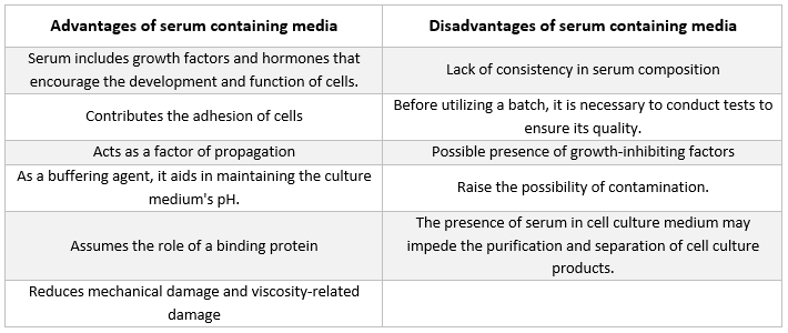
Loại chất bổ sung thường gặp nhất trong môi trường dùng để nuôi cấy tế bào động vật là huyết thanh bò thai. Chất này được thêm vào môi trường nuôi cấy như một chất bổ sung giá rẻ nhằm đạt được điều kiện phát triển tốt nhất có thể. Ngoài vai trò là chất vận chuyển hoặc chất chelat hóa cho các chất dinh dưỡng không ổn định hoặc không tan trong nước, hormone và các yếu tố tăng trưởng, chất ức chế protease và các chất khác, huyết thanh còn liên kết và trung hòa các phân tử có hại.

Môi trường không có huyết thanh

Sự hiện diện của huyết thanh trong môi trường nuôi cấy có một số nhược điểm và có khả năng gây ra những sai sót lớn trong việc giải thích kết quả nghiên cứu miễn dịch [2, 3]. Đã có nhiều loại môi trường nuôi cấy không chứa huyết thanh khác nhau được tạo ra [4, 5]. Các môi trường này thường được thiết kế đặc biệt để hỗ trợ nuôi cấy một loại tế bào cụ thể, chẳng hạn như Knockout Serum Replacement và Knockout DMEM của Thermo Fisher Scientific, cùng với môi trường mTESR của Stem Cell Technologies [6], dành cho tế bào gốc [7].

Ngoài ra, các môi trường này còn chứa một lượng xác định các yếu tố tăng trưởng tinh khiết, lipoprotein và các protein khác, vốn thường được cung cấp bởi huyết thanh [8]. Các môi trường này thường được gọi là “môi trường nuôi cấy xác định” vì các thành phần cấu thành chúng đã được hiểu rõ.

Môi trường nuôi cấy được xác định về mặt hóa học

Các môi trường này bao gồm các thành phần vô cơ và hữu cơ siêu tinh khiết, không bị nhiễm bất kỳ loại tạp chất nào. Chúng cũng có thể bao gồm các chất bổ sung protein tinh khiết, chẳng hạn như các yếu tố tăng trưởng.

 Việc biến đổi gen của vi khuẩn hoặc nấm men, cùng với việc bổ sung các axit béo, vitamin, cholesterol và axit amin cụ thể, dẫn đến việc sản xuất các thành phần của chúng [9].

Môi trường không chứa protein

Môi trường không chứa protein là những môi trường hoàn toàn không chứa protein mà chỉ bao gồm các thành phần phi protein. So với môi trường có bổ sung huyết thanh, việc sử dụng môi trường không bổ sung protein thúc đẩy sự tăng sinh tế bào và biểu hiện protein mạnh mẽ hơn, đồng thời giúp việc tinh chế bất kỳ sản phẩm nào được tạo ra trong quá trình hạ lưu trở nên dễ dàng hơn [10-12]. Protein không được bao gồm trong các công thức như MEM và RPMI-1640. Tuy nhiên, có thể bổ sung protein nếu cần thiết.

Môi trường nuôi cấy và các thành phần cơ bản của nó

Môi trường nuôi cấy thương mại có thể được mua dưới dạng bột hoặc chất lỏng và thường chứa nhiều chất dinh dưỡng khác nhau như axit amin, glucose, muối, vitamin và các chất bổ sung dinh dưỡng khác. 

Nhu cầu về các thành phần này khác nhau đối với từng dòng tế bào, và những sự khác biệt này là nguyên nhân dẫn đến sự đa dạng về công thức môi trường nuôi cấy. Mỗi thành phần đảm nhiệm một chức năng nhất định, sẽ được trình bày trong các đoạn sau:

Hệ thống đệm

Để duy trì điều kiện sinh trưởng tối ưu, pH phải được kiểm soát, thường được thực hiện bằng một trong hai hệ thống đệm sau:

Hệ thống đệm tự nhiên

Tỷ lệ CO2/H2CO3 trong khí quyển bằng với tỷ lệ trong môi trường nuôi cấy, tạo ra một cơ chế đệm tự nhiên. Để duy trì cơ chế đệm tự nhiên này, các mẫu nuôi cấy phải được giữ trong môi trường không khí có nồng độ CO2 từ 5-10%, điều này thường được thực hiện bằng cách sử dụng tủ ấp CO2. Một trong những ưu điểm lớn nhất của việc sử dụng chất đệm tự nhiên là chi phí rẻ và an toàn.

HEPES

Đệm hóa học sử dụng ion lưỡng tính HEPES có khả năng đệm cao hơn trong phạm vi pH 7,2-7,4 và không cần môi trường khí được điều chỉnh. Đối với một số loại tế bào cụ thể, liều lượng HEPES cao hơn có thể gây hại. Môi trường nuôi cấy có chứa HEPES cũng dễ bị ảnh hưởng bởi tác động quang độc của ánh sáng huỳnh quang hơn nhiều [13].

Phenol Red

Chất chỉ thị pH phenol red thường được bổ sung vào môi trường nuôi cấy thương mại, cho phép theo dõi liên tục giá trị pH. Khi tế bào phát triển, các chất chuyển hóa do chúng sản sinh ra gây ra sự thay đổi pH và do đó làm thay đổi màu sắc của môi trường nuôi cấy. Phenol Red có tác động kép lên màu sắc của môi trường nuôi cấy: chuyển sang màu vàng ở pH axit và màu tím ở pH kiềm. pH 7,4 – giá trị tối ưu cho nuôi cấy tế bào – khiến môi trường nuôi cấy có màu đỏ huỳnh quang.

Tuy nhiên, phenol red có một số nhược điểm: Thứ nhất, phenol red có khả năng mô phỏng hoạt động của một số hormone steroid, chủ yếu là estrogen [14]. Do đó, khi nghiên cứu các tế bào nhạy cảm với estrogen như mô vú, nên sử dụng môi trường nuôi cấy không chứa phenol red. Sự cân bằng natri-kali bị phá vỡ do sự hiện diện của phenol red trong một số công thức không có huyết thanh. Việc thêm huyết thanh hoặc hormone tuyến yên bò vào môi trường nuôi cấy có thể khắc phục tác động này [15]. Thứ ba, việc phát hiện trong các thí nghiệm đo lưu lượng tế bào bị cản trở do sự hiện diện của phenol red.

Muối vô cơ

Môi trường nuôi cấy chứa muối vô cơ, chẳng hạn như ion natri, kali và canxi, giúp duy trì cân bằng thẩm thấu và điều chỉnh điện thế màng tế bào.

Axit amin

Vì axit amin là thành phần cơ bản của protein, chúng là thành phần thiết yếu của mọi môi trường nuôi cấy tế bào từng được phát minh. Do tế bào không thể tự sản xuất một số axit amin nhất định, nên điều quan trọng là môi trường nuôi cấy phải chứa các axit amin thiết yếu. Chúng cần thiết cho sự nhân lên của tế bào, và nồng độ của chúng quyết định mật độ tế bào tối đa có thể đạt được. Đặc biệt, L-glutamine, một axit amin thiết yếu, có vai trò đặc biệt quan trọng.

L-glutamine đóng vai trò là nguồn năng lượng thứ cấp cho quá trình chuyển hóa và cung cấp nitơ cho việc sản xuất NAD, NADPH và các nucleotide. Do L-glutamine là một axit amin không ổn định, theo thời gian sẽ chuyển hóa thành dạng mà tế bào không thể sử dụng được, nên nó phải được bổ sung vào môi trường nuôi cấy.

Ngoài ra, các axit amin không thiết yếu có thể được bổ sung vào môi trường nuôi cấy để bù đắp lượng đã bị tiêu hao trong quá trình sinh trưởng. Sự sinh trưởng của tế bào được thúc đẩy và khả năng sống sót của chúng được tăng cường khi môi trường nuôi cấy được bổ sung các axit amin không thiết yếu.

Carbohydrate

Carbohydrate dưới dạng đường là nguồn năng lượng chính. Ngoài các loại đường phổ biến như glucose và galactose, nhiều môi trường nuôi cấy còn chứa maltose và fructose.

Protein và peptit

Albumin, transferrin và fibronectin là các protein và peptit được sử dụng phổ biến nhất. Chúng đặc biệt quan trọng trong các môi trường nuôi cấy không chứa huyết thanh. Albumin, transferrin, aprotinin, fetuin và fibronectin là một số protein có thể tìm thấy trong huyết thanh, vốn là nguồn cung cấp protein dồi dào.

Albumin là protein chính có trong máu, có chức năng liên kết và vận chuyển các chất khác nhau, bao gồm nước, muối, axit béo tự do, hormone và vitamin, giữa các cơ quan và tế bào khác nhau. Khả năng liên kết với các chất hóa học của albumin khiến nó trở thành một ứng cử viên hiệu quả để loại bỏ các hợp chất có hại khỏi môi trường nuôi cấy tế bào.

Aprotinin là chất bảo vệ trong hệ thống nuôi cấy tế bào, do nó ổn định ở pH trung tính và axit, cũng như chịu được nhiệt độ cao và sự phân hủy có thể do các enzym proteolytic gây ra. Chất này có khả năng ức chế một số protease serine, bao gồm trypsin và các loại khác.

Fetuin là một glycoprotein có thể được phát hiện với lượng cao hơn trong huyết thanh của thai nhi và động vật sơ sinh so với huyết thanh của động vật trưởng thành. Ngoài ra, nó còn hoạt động như một chất ức chế protease serine. Protein fibronectin là thành phần thiết yếu trong quá trình bám dính tế bào. Transferrin là một loại protein vận chuyển sắt và chịu trách nhiệm cung cấp sắt cho màng tế bào.

Axit béo và lipid

Chúng đóng vai trò quan trọng trong môi trường nuôi cấy không có huyết thanh khi huyết thanh vắng mặt.

Vitamin

Nhiều loại vitamin là cần thiết cho sự phát triển và sinh sôi của tế bào. Tế bào không thể tự sản xuất vitamin với lượng đủ nên vitamin là chất bổ sung dinh dưỡng thiết yếu trong nuôi cấy mô.

Trong nuôi cấy tế bào, huyết thanh là nguồn vitamin chính; tuy nhiên, môi trường nuôi cấy cũng được bổ sung các loại vitamin khác nhau để phù hợp với từng loại tế bào cụ thể. Thông thường, các vitamin nhóm B được sử dụng để kích thích sự tăng trưởng.

Các nguyên tố vi lượng

Các nguyên tố hóa học như đồng, kẽm, selen và các chất trung gian của axit tricarboxylic được gọi là nguyên tố vi lượng. Nguyên tố vi lượng thường được bổ sung vào môi trường nuôi cấy không chứa huyết thanh để thay thế những nguyên tố thường có trong huyết thanh. Những nguyên tố này là các thành phần hóa học quan trọng cần thiết cho sự phát triển khỏe mạnh của tế bào. Nhiều phản ứng sinh hóa phụ thuộc vào các vi chất dinh dưỡng nhất định, chẳng hạn như hoạt động của enzyme.

Chất bổ sung môi trường nuôi cấy

Môi trường nuôi cấy hoàn chỉnh được đề xuất cho một số dòng tế bào nhất định cần có các thành phần bổ sung không có trong môi trường cơ bản và huyết thanh. Các chất bổ sung này hỗ trợ sự phát triển của tế bào và chức năng trao đổi chất thích hợp.

Mặc dù hormone, yếu tố tăng trưởng và các phân tử tín hiệu là yếu tố thiết yếu cho sự nhân lên thích hợp của các dòng tế bào cụ thể, nhưng luôn cần tuân thủ các biện pháp phòng ngừa sau: Vì việc bổ sung các chất này có thể làm thay đổi độ thẩm thấu của môi trường nuôi cấy hoàn chỉnh, từ đó có thể ức chế sự phát triển của tế bào, nên luôn cần kiểm tra lại độ thẩm thấu sau khi bổ sung. Đối với phần lớn các dòng tế bào, độ thẩm thấu tối ưu dao động trong khoảng từ 260 đến 320 mOSM/kg.

Kháng sinh

Kháng sinh thường được sử dụng để ức chế sự phát triển của các tác nhân gây ô nhiễm vi khuẩn và nấm [16], mặc dù chúng không phải là yếu tố thiết yếu cho sự phát triển của tế bào. Do kháng sinh có thể che giấu sự nhiễm bẩn bởi mycoplasma và vi khuẩn kháng thuốc, việc sử dụng thường xuyên kháng sinh không được khuyến nghị trong nuôi cấy tế bào [17, 18].

Ngoài ra, kháng sinh có thể làm rối loạn quá trình trao đổi chất của các tế bào quá mẫn cảm. Các hỗn hợp penicillin-streptomycin do MilliporeSigma và Life Technologies sản xuất thường được sử dụng. Plasmocin đã được sử dụng trong nuôi cấy các dòng tế bào u thần kinh đệm TS603, TS516 và BT260 [19], và đã được chứng minh là có hiệu quả trong việc loại bỏ nhiễm mycoplasma (20).

Huyết thanh

Albumin, các yếu tố tăng trưởng và các chất ức chế tăng trưởng đều có trong huyết thanh. Huyết thanh là một trong những thành phần quan trọng nhất của môi trường nuôi cấy tế bào vì nó cung cấp axit amin, protein, vitamin (đặc biệt là các vitamin tan trong chất béo như A, D, E và K), carbohydrate, lipid, hormone, các yếu tố tăng trưởng, khoáng chất và các nguyên tố vi lượng.

Huyết thanh từ nguồn bò thai và bê thường được sử dụng để thúc đẩy sự phát triển của tế bào nuôi cấy. Huyết thanh bò thai là nguồn cung cấp dồi dào các yếu tố tăng trưởng và phù hợp cho việc nhân bản tế bào cũng như phát triển các tế bào nhạy cảm. Do khả năng thúc đẩy tăng trưởng bị suy giảm, huyết thanh bê được sử dụng trong các thí nghiệm ức chế tiếp xúc. Môi trường nuôi cấy thông thường thường chứa từ 2% đến 10% huyết thanh. Việc bổ sung huyết thanh vào môi trường nuôi cấy phục vụ các mục đích sau [21]:

  • Huyết thanh cung cấp các chất dinh dưỡng thiết yếu cho tế bào (cả ở dạng dung dịch và gắn với protein).

  • Huyết thanh chứa một số yếu tố tăng trưởng và hormone tham gia vào quá trình thúc đẩy tăng trưởng và hoạt động chuyên biệt của tế bào.

  • Nó cung cấp nhiều protein liên kết, như albumin và transferrin, giúp vận chuyển các chất hóa học khác vào tế bào. Ví dụ, albumin vận chuyển chất béo, vitamin, hormone, v.v. vào tế bào.

  • Nó cũng cung cấp các protein, chẳng hạn như fibronectin, giúp tăng khả năng bám dính của tế bào vào chất nền. Ngoài ra, nó còn tạo ra các yếu tố lan tỏa giúp tế bào mở rộng trước khi phân chia.

  • Nó cung cấp các chất ức chế protease giúp ngăn chặn quá trình phân giải protein trong tế bào.

  • Nó cũng chứa các khoáng chất như Na+, K+, Zn2+ và Fe2+.

  • Nó làm tăng độ nhớt của môi trường nuôi cấy, từ đó bảo vệ tế bào khỏi tổn thương cơ học trong quá trình khuấy trộn khi nuôi cấy tế bào trong dung dịch lơ lửng.

  • Nó cũng đóng vai trò như một chất đệm.
Tài liệu tham khảo

[1] Morgan J, Morton H, Parker R. Dinh dưỡng của tế bào động vật trong nuôi cấy mô; các nghiên cứu ban đầu về môi trường nuôi cấy tổng hợp. Proc Soc Exp Biol Med. 1950;73:1-8

[2] Kerbel R, Blakeslee D. Sự hấp phụ nhanh chóng của một thành phần huyết thanh bò thai bởi các tế bào động vật có vú trong nuôi cấy. Một nguồn tiềm ẩn gây ra hiện tượng giả trong các nghiên cứu về huyết thanh kháng thể đối với các kháng nguyên đặc hiệu tế bào. Immunology. 1976;31:881-91

[3] Sula K, Draber P, Nouza K. Việc bổ sung huyết thanh vào môi trường dùng để chuẩn bị huyền phù tế bào là một nguồn có thể gây ra sai lệch trong các phản ứng miễn dịch tế bào được nghiên cứu thông qua thử nghiệm hạch bạch huyết vùng gối. Tạp chí Di truyền Miễn dịch. 1980;7:483-9

[4] Mariani E, Mariani A, Monaco M, Lalli E, Vitale M, Facchini A. Môi trường nuôi cấy không chứa huyết thanh thương mại: sự phát triển của tế bào lai và sản xuất kháng thể đơn dòng. Tạp chí Phương pháp Miễn dịch học. 1991;145:175-83

[5] Barnes D, Sato G. Các phương pháp nuôi cấy tế bào trong môi trường không chứa huyết thanh. Anal Biochem. 1980;102:255-70

[6] Yu H, Lu S, Gasior K, Singh D, Vazquez Sanchez S, Tapia O, và cộng sự. Các protein chaperone HSP70 vận chuyển TDP-43 không chứa RNA vào các vỏ cầu lỏng nội nhân có tính dị hướng. Science. 2021;371:

[7] Meharena H, Marco A, Dileep V, Lockshin E, Akatsu G, Mullahoo J, và cộng sự. Quá trình lão hóa do hội chứng Down gây ra làm rối loạn cấu trúc nhân của các tế bào tiền thân thần kinh. Cell Stem Cell. 2022;29:116-130.e7

[8] Iscove N, Melchers F. Thay thế hoàn toàn huyết thanh bằng albumin, transferrin và lipid đậu nành trong môi trường nuôi cấy các tế bào lympho B phản ứng với lipopolysaccharide. J Exp Med. 1978;147:923-33

[9] Stoll T, Muhlethaler K, von Stockar U, Marison I. Cải tiến có hệ thống môi trường nuôi cấy không chứa protein và được định nghĩa hóa học dành cho sự phát triển của tế bào hybridoma và sản xuất kháng thể đơn dòng. J Biotechnol. 1996;45:111-23

[10] Darfler F. Môi trường không chứa protein để nuôi cấy tế bào lai và các tế bào khác của hệ miễn dịch. In Vitro Cell Dev Biol. 1990;26:769-78

[11] Barnes D, Sato G. Nuôi cấy tế bào không huyết thanh: một phương pháp thống nhất. Cell. 1980;22:649-55

[12] Hamilton W, Ham R. Sự phát triển dòng tế bào chuột hamster Trung Quốc trong môi trường không chứa protein. In Vitro. 1977;13:537-47

[13] Zigler J, Lepe Zuniga J, Vistica B, Gery I. Phân tích tác dụng độc tế bào của môi trường nuôi cấy chứa HEPES khi tiếp xúc với ánh sáng. In Vitro Cell Dev Biol. 1985;21:282-7

[14] Berthois Y, Katzenellenbogen J, Katzenellenbogen B. Chất Phenol red trong môi trường nuôi cấy mô là một estrogen yếu: những hàm ý liên quan đến việc nghiên cứu các tế bào nhạy cảm với estrogen trong nuôi cấy. Proc Natl Acad Sci U S A. 1986;83:2496-500

[15] Karmiol S. Phát triển môi trường nuôi cấy không có huyết thanh. Trong: Master JRW, biên tập. Animal Cell culture, ấn bản thứ 3. Oxford: Oxford University Press; 2000.

[16] Perlman D. Việc sử dụng kháng sinh trong môi trường nuôi cấy tế bào. Methods Enzymol. 1979;58:110-6

[17] McGarrity G. Sự lây lan và kiểm soát nhiễm trùng mycoplasma trong nuôi cấy tế bào. In Vitro. 1976;12:643-8

[18] Masters J, Stacey G. Thay đổi môi trường nuôi cấy và nhân giống dòng tế bào. Nat Protoc. 2007;2:2276-84

[19] Chakraborty A, Laukka T, Myllykoski M, Ringel A, Booker M, Tolstorukov M, và cộng sự. Enzym demethylase histone KDM6A trực tiếp cảm nhận oxy để điều chỉnh cấu trúc chromatin và số phận tế bào. Science. 2019;363:1217-1222

[20] Molla Kazemiha V, Azari S, Amanzadeh A, Bonakdar S, Shojaei Moghadam M, Habibi Anbouhi M, và cộng sự. Hiệu quả của Plasmocin™ trên các dòng tế bào động vật có vú bị nhiễm mollicutes so với các loại kháng sinh thường được sử dụng trong nuôi cấy tế bào: kinh nghiệm tại địa phương. Cytotechnology. 2011;63:609-20

[21] Kragh Hansen U. Các khía cạnh phân tử của sự liên kết ligand với albumin huyết thanh. Pharmacol Rev. 1981;33:17-53